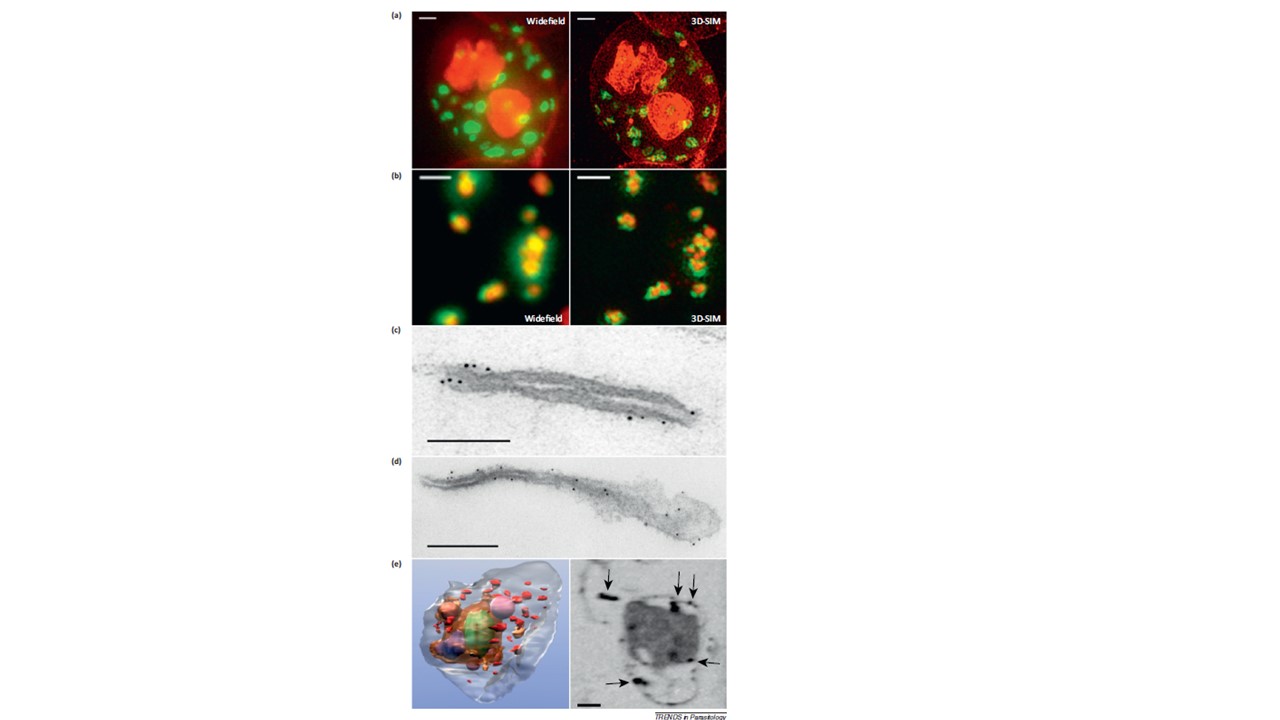
Microscopy methods for localizing Plasmodium falciparum Maurer’s cleft-associated proteins. (a) Plasmodium falciparum transfectants expressing REX1-GFP (green) costained with a membrane stain (red) and imaged using conventional widefield fluorescence microscopy and 3D structured illumination microscopy (3D-SIM). Scale bars = 1 mm. (b) Immunofluorescence microscopy of P. falciparum REX1-GFP transfectants labeled with anti-GFP (green) and anti-MAHRP1 (red) antibodies. The sample was imaged using conventional widefield fluorescence microscopy and 3D-SIM. Scale bars = 1 mm. (c, d) Immunoelectron microscopy. Red blood cells (RBCs) infected with trophozoite stage REX1-GFP transfectants [(c) scale bar = 100 nm)] or MAHRP1-GFP transfectants [(d) (scale bar = 200 nm)] were permeabilized with Equinatoxin II and labeled with anti-GFP antiserum followed by 6 nm gold-labeled protein A. (e) Left hand panel: rendered cryo-X-ray tomogram of an Equinatoxin II permeabilized trophozoite-infected RBC [57]. The surface of the trophozoite is rendered in brown, the nucleus in green, the digestive vacuole in purple. Hemoglobin containing vesicles in the RBC cytoplasm are rendered in magenta and Maurer’s clefts in red. Right hand panel: cryo-X-ray micrograph of an infected RBC. MAHRP1-GFP transfectant-infected RBCs were permeabilized and labeled with mouse anti-GFP antiserum followed by 6 nm gold-labeled protein A. The size of the gold particles was increased by silver enhancement. Dark deposits in the RBC cytoplasm (arrowheads) reveal labeling of the Maurer’s clefts. Scale bars = 500 nm. Woodcroft BJ, McMillan PJ, Dekiwadia C, Tilley L, Ralph SA. Determination of protein subcellular localization in apicomplexan parasites. Trends Parasitol. 2012 Dec;28(12):546-54. PMID: 22995720ÿÿ