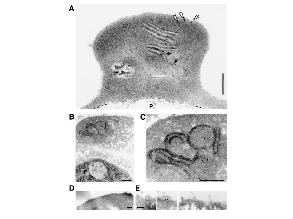
Electron microscopic immunolocalization of PfEMP1 onultrathin sections of erythrocytes infected with P. falciparum using secondary antibodies coupled to 12 nm colloidal gold particles. (A) Electron micrograph showing anti-PfEMP1 labeling of long slender, stacked tubular membranes. The white arrowheads mark an oblique sectioned membrane stack that appears to extend to the erythrocyte surface. This membrane is gold labeled beneath the erythrocyte plasma membrane (white arrow). Black arrows show gold labeling on cross sectioned tubular profiles. The broken line marks the border between the parasite (P) and the erythrocyte cytoplasm. (B, C). Electron micrographs of ultrathin sections showing anti-PfEMP1 labeling of an extended, membranous network with whorls and vesicle-like profiles. Gold particles were also detectable over the parasite (P). (C) shows the boxed area marked in (B) at higher magnification. (D) Electron micrograph showing that P. falciparum clone HB3 trophozoite-infected erythrocytes possess knobs. Cells were fixed with glutardialdehyde and osmium tetroxide. (E) Electron micrographs of the surface of the same cell shown in (B) revealing anti-PfEMP1 labeling of the host erythrocyte surface in structures resembling knobs. Bars, 200 nm (A-D); 100 nm (E).
Wickert H, Wissing F, Andrews KT, Stich A, Krohne G, Lanzer M. Evidence for trafficking of PfEMP1 to the surface of P. falciparum-infected erythrocytes via a complex membrane network. Eur J Cell Biol. 2003 82:271-84.
