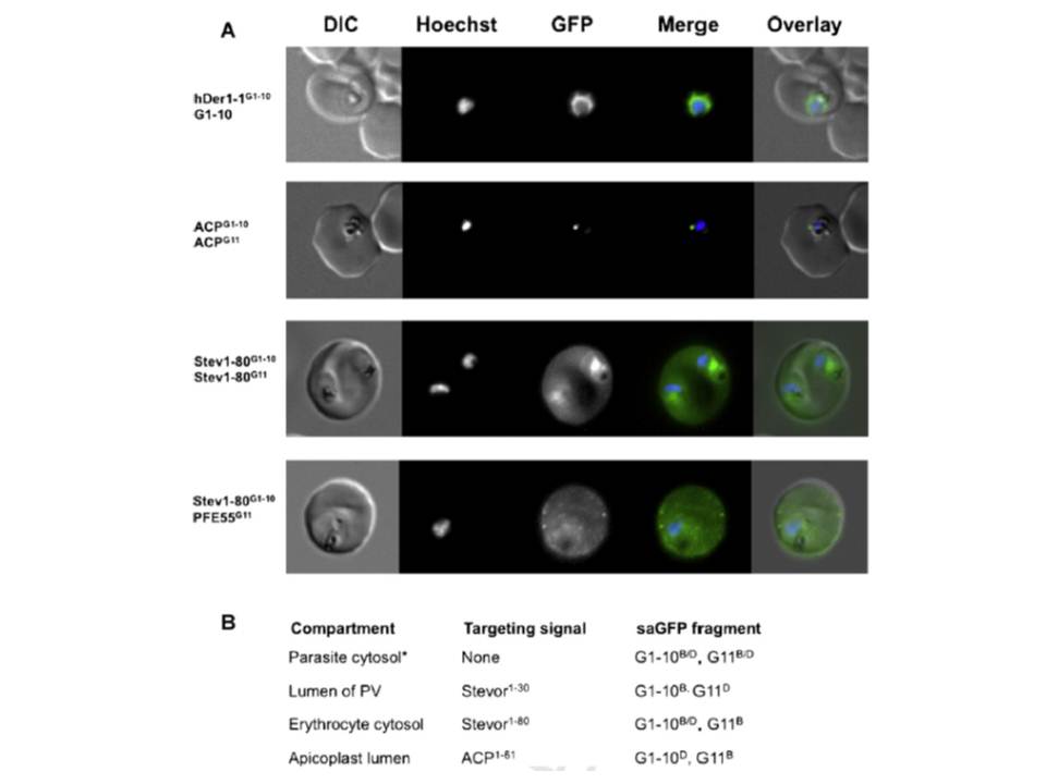
(A) Live cell imaging of co-transfectants expressing saGFP (self-assembling split GFP) fragments in various cellular compartments. In merge and overlay: green, GFP; blue, Hoechst. (B) Vectors available for analysis of cellular compartmentalisation using saGFP. Numbers in targeting sequence refer to N-terminal amino acids. B, Blasticidin-S-deaminase resistance cassette; D, hDHFR resistance cassette. *Can be used to generate fusions as multiple cloning sites situated in front of saGFP coding sequence.
Külzer S, Petersen W, Baser A, Mandel K, Przyborski JM. Use of self-assembling GFP to determine protein topology and compartmentalisation in the Plasmodium falciparum-infected erythrocyte. Mol Biochem Parasitol. 2012 187(2):87-90.
PubMed Article: Use of self-assembling GFP to determine protein topology and compartmentalisation in the Plasmodium falciparum-infected erythrocyte
Other associated proteins
| PFID | Formal Annotation |
|---|---|
| PF3D7_0208500 | acyl carrier protein |
| PF3D7_0501100 | co-chaperone j domain protein jdp |
| PF3D7_1452300 | DER1-like protein |
