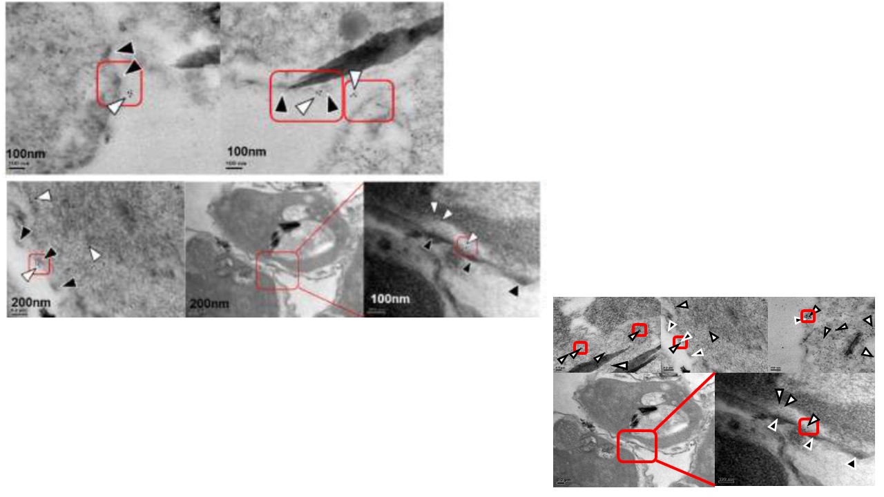
Upper and middle pannels: Clustering of surface exposed STEVOR in proximity to knobs. Right upper panel: Representative images from pre embedded TEM imaging of ultra-thin slices (~90nm) of late stage 5A iRBCs. Gold particle clusters can be observed in proximity to surface knobs of cells. White arrows: 10nm Au particles; black arrows: knobs. Right lower panel: Representative images from post embedded TEM imaging of ultra-thin slices (~90nm) of late stage 5A iRBCs. Third image in the row shows the zoom in view of the red highlighted region in second image. Gold tagged STEVOR can be observed in different parts of the cell and in close proximity to surface knobs in near surface region. Scale bars have been indicated in images. Images from pre embedding immunoTEM of iRBC showed clusters of gold nanoparticles on the extracellular surface of cells and post embedding immunoTEM of fixed iRBC revealed localizations of Au nanoparticles in different parts of the cell - Maurer's Clefts, RBC membrane and cell surface knobs in near surface region. Lower right panel: Representative images of 5A, A4(tr-I) and A4(tr-II) cells upon post-embedding immunostaining with anti-S1 serum followed by 10nm gold conjugated secondary antibodies. Clusters of gold nanoparticles can be seen in different parts of the cell (highlighted in red boxes). White arrows indicate STEVOR; black arrows indicate a knob. A small fraction of gold particles are also observed being localized on the outer surface of cells. Many of these gold particles (both in intracellular and outer surface regions) can be seen in close proximity of knobs. First image in row 2 shows the full view of an example cell with a zoom in view of the red highlighted box in the second image of the same row.
Singh H, Madnani K, Lim YB, Cao J, Preiser PR, Lim CT. Expression dynamics & physiologically relevant functional study of STEVOR in asexual stages of Plasmodium falciparum infection. Cell Microbiol. 2016 Dec 28.
