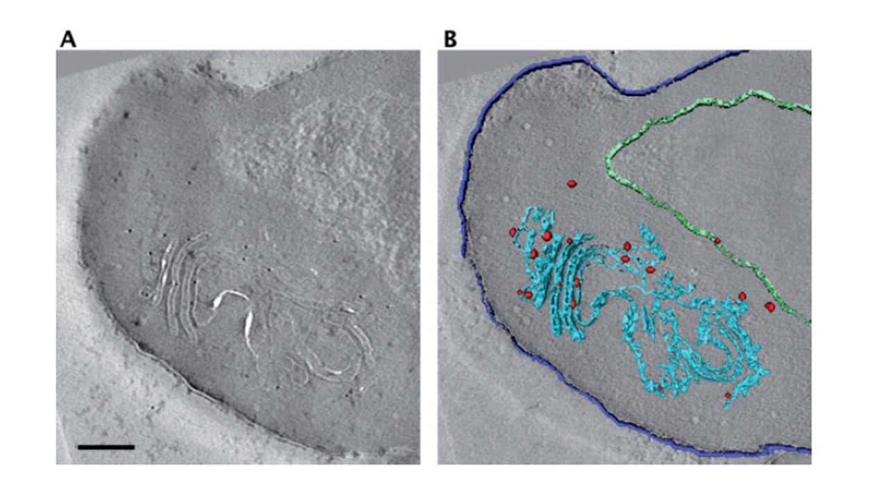
‘Tokuyasu’ section of infected erythrocyte, immunolabeled against STEVOR. (A) Section through a tomogram showing morphology of Maurer’s cleft and STEVOR labeling. (B) Surface rendered view of the area in (A). Blue, Maurer’s cleft; dark blue, outline of erythrocyte plasma membrane; green, outline of parasitophorous vacuole; red, positions of gold markers. Thickness of the section: 200 nm. Scale bar: 200 nm. Tokuyasu sections and freeze-substituted sections are better suited for immuno-EM studies. Using parasites expressing a STEVOR-GFP fusion protein, we were able to localize the protein to the Maurer’s clefts in Tokuyasu sections (A and B). However, owing to the size of an antibody and the gold label the resolution is approximately 10–15 nm. Thus, for the Maurer’s clefts stacks, it would be difficult to determine whether the antigenic sites are located on the membranes or on connectors between the membranes, without statistical analysis.
Henrich P, Kilian N, Lanzer M, Cyrklaff M. 3-D analysis of the Plasmodium falciparum Maurer's clefts using different electron tomographic approaches. Biotechnol J. 2009 4(6):888-94.
