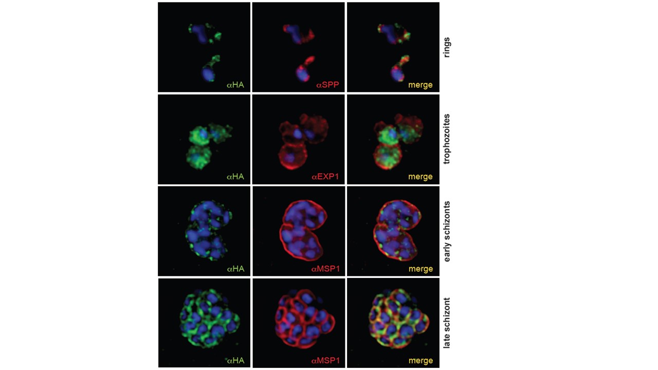
Immunofluorescence analysis of PfPLSCR-HA expressing parasites. The upper panel depicts two ring-stage parasites and co-labelling with anti-SPP (signal peptide peptidase) antibodies as an ER marker. Anti-EXP1 (exported protein 1) delineates the PVM surrounding three trophozoites in a multiply infected red blood cell. Lower two panels show deconvolved co-labelling with antibodies against MSP1 (merozoite surface protein 1) in late blood stage parasites. PfPLSCR-HA co-localises with MSP1 at the plasma membrane in late schizonts. Parasite nuclei are stained with DAPI (blue). Scale bar = 1 mM. Immunofluorescence analysis revealed the localisation of the epitope-tagged protein to the periphery of individual merozoites in late schizonts as suggested by co-localisation with the merozoite surface protein MSP1. The protein also localised to a perinuclear region in the early ring stages as evident by partial co-localisation with the ER-resident protein signal peptide peptidase, and redistributed to the cytoplasm and vesicular foci in early trophozoite and schizont stages
Haase S, Condron M, Miller D, Cherkaoui D, Jordan S, Gulbis JM, Baum J. Identification and characterisation of a phospholipid scramblase in the malaria parasite Plasmodium falciparum. Mol Biochem Parasitol. 2021 8:111374.
Other associated proteins
| PFID | Formal Annotation |
|---|---|
| PF3D7_0930300 | merozoite surface protein 1 |
| PF3D7_1121600 | parasitophorous vacuole membrane antigen QF 116 exported protein 1 circumsporozoite-related antigen |
| PF3D7_1457000 | signal peptide peptidase |