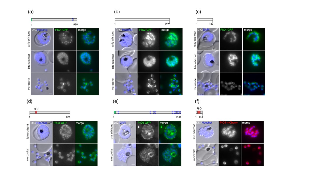
Localization of PhIL1 interacting candidates (PIC) using endogenous GFP tagging. (a) PIC1, (b) PIC2 (c) PIC4 (d) PIC3, (e): PIC6: On top a schematic representation of the protein (protein length indicated as number of amino acids) with putative protein domains (blue, transmembrane domain; green, signal peptide; red, Zinc Finger domain (ZFD) or RBD (RNA binding domain). Localization of PIC-GFP fusion proteins in schizonts and free merozoites are shown in the middle panel, the PCR-based confirmations of the correct insertion of GFP-encoding integration plasmid into the targeted loci on the bottom. Ladder size indicated in base pairs (bp). gDNA from parental 3D7 was used as control. (f) Localization of PIC5-mCherry fusion proteins episomally expressed under the control of the late-stage promoter ama1 in schizonts and free merozoites. Nuclei stained with Hoechst-33,342 or DAPI. Zoom factor: 400%. Scale bar 2 μm. KI, knock-in
Wichers JS, Wunderlich J, Heincke D, Pazicky S, Strauss J, Schmitt M, Kimmel J, Wilcke L, Scharf S, von Thien H, Burda PC, Spielmann T, Löw C, Filarsky M, Bachmann A, Gilberger TW. Identification of novel inner membrane complex and apical annuli proteins of the malaria parasite Plasmodium falciparum. Cell Microbiol. 2021 8:e13341.
Other associated proteins
| PFID | Formal Annotation |
|---|---|
| PF3D7_0308300 | phil1-interacting candidate pic4 |
| PF3D7_0415800 | phil1-interacting candidate pic3 |
| PF3D7_0530300 | phil1-interacting candidate pic6 |
| PF3D7_0822900 | phil1-interacting candidate pic2 |
| PF3D7_1229300 | phil1-interacting candidate pic1 |
| PF3D7_1310700 | PhIL1-interacting protein PhIP |