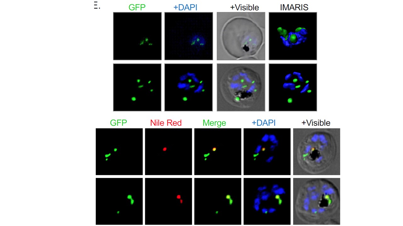
Upper panel. Fluorescent microscopic images of transgenic parasites. Fusion protein signal was observed in vesicular structures in transgenic parasites. The parasite nuclei were stained with DAPI and parasites were visualized by a confocal laser scanning microscope. The GFP signal in these parasites was observed in vesicular structures in the parasite cytosol as distinct foci/vesicle during trophozoite stages, whereas during late-trophozoite and schizont stages, the labelling was observed in large vesicular structure present juxtaposed to the food vacuole (upper). Lower panel. PfLPL20 associate with a neutral lipid storage body near the food vacuole. Fluorescence images of trophozoites stage transgenic parasites expressing PfLPL20-GFP stained with Nile Red, a neutral lipid staining dye. The large GFP labelled structure close to the food vacuole (having dark hemozoin). Further, staining with Nile red showed that these vesicular structures are neutral lipid-rich body; Nile red staining showed overlap with the endogenously tagged PfLPL20-GFP protein in these structures.
Sheokand PK, Narwal M, Thakur V, Mohmmed A. GlmS mediated knock-down of a phospholipase expedite alternate pathway to generate phosphocholine required for phosphatidylcholine synthesis in Plasmodium falciparum. Biochem J. 2021 Jun 16:BCJ20200549.