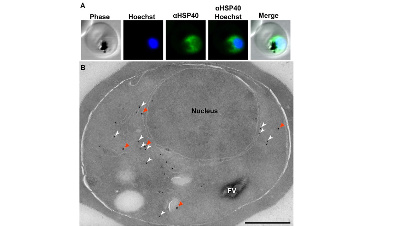
Localization of HSP40 in P. falciparum. (A) Immunofluorescence confocal microscopy of trophozoite, stained with anti-HSP40 (1:5,000) and Hoechst 33258 nuclear stain. HSP40 appears cytosolic. (B) Electron micrograph of immunolabeling: primary, rabbit anti-HSP40 (1:250), mouse anti-PDI (1:100); secondary, goat anti-rabbit IgG 18nm colloidal gold, goat anti-mouse 12 nm. HSP40 (orange arrowheads) looks cytosolic in the parasites, with some apparent membrane association. A portion of HSP40 colocalizes with PDI, an established ER marker (white arrowheads). Scale, 500 nm. HSP40 localizes to the parasite cytosol in trophozoite-stage parasites and is not detected in the host cell (A). Immuno-electron microscopy (immuno-EM) was performed to further characterize the subcellular localization of HSP40. We found that HSP40 is distributed throughout the cytosol and excluded from the nucleus and host cell cytoplasm (B). parasites were labeled with anti-protein disulfide isomerase (anti-PDI), an established ER marker. A subset of HSP40 (48.7% of all labeling) is localized alongside PDI in the ER (B). These data indicate that HSP40 is both cytosolic and ER localized.
Mathews ES, Jezewski AJ, Odom John AR. Protein Prenylation and Hsp40 in Thermotolerance of Plasmodium falciparum Malaria Parasites. mBio. 2021 29:e0076021
Other associated proteins
| PFID | Formal Annotation |
|---|---|
| PF3D7_1437900 | HSP40, subfamily A |