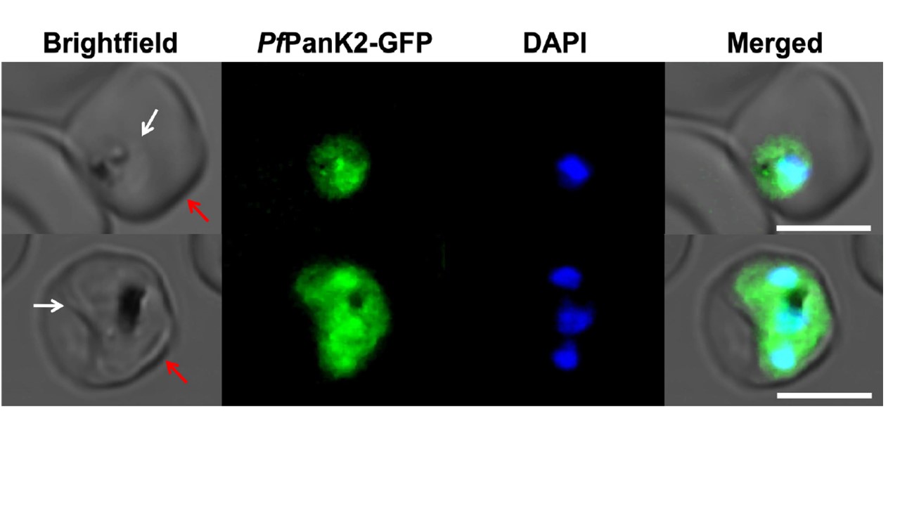
Confocal micrographs showing the subcellular location of PfPanK2-GFP within trophozoite/schizont-stage P. falciparum-infected erythrocytes. The nuclei of the parasites are stained with DAPI. From left to right: Brightfield, GFP-fluorescence, DAPI-fluorescence, and merged images. Arrows indicate the plasma membranes of the erythrocyte (red) or the parasite (white). Scale bars represent 5 μm. We episomally expressed PfPanK2-GFP in asexual blood stage P. falciparum parasites and found that PfPanK2-GFP is localised throughout the parasite cytosol and is not excluded from the nucleus.
Tjhin ET, Howieson VM, Spry C, van Dooren GG, Saliba KJ. A novel heteromeric pantothenate kinase complex in apicomplexan parasites. PLoS Pathog. 2021 17(7):e1009797.