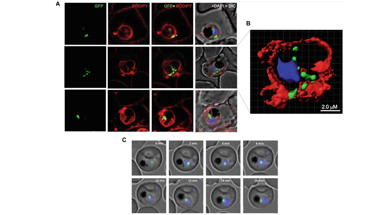
PfLPL1 resides in vesicles which associate with parasite membrane and with food-vacuole. a Fluorescence images showing labelling of membranes in P. falciparum infected RBCs and localization of PfLPL1. Trophozoite stage transgenic parasites expressing PfLPL1-GFP-DDD were stained with BODIPY-TR ceramide (red); the parasite nuclei were stained with DAPI (blue) and visualized by confocal laser scanning microscope. Small GFP foci of the PfLPL1-GFP-DDD fusion protein was observed near parasite boundary (panel 1, marked with arrowhead); these foci showed closed association with fluorescence ed parasite membrane. In some parasites, the GFP vesicles are seen in parasite cytosol (panel 2) and in close association with food-vacuole (panels 2 and 3 marked with arrowhead). b A three-dimensional reconstruction of series of Z-stack images (corresponding to panel 2 in a) using IMARIS software. Small GFP vesicles are present juxtaposed to the parasite membrane, in the parasite cytosol and close to the food-vacuole. c Time-lapse microscopy of PfLPL1-GFP-DDD expressing transgenic parasites showing localization and migration GFP-labelled vesicles. Sequential images of a trophozoite stage parasite over a time interval of 16 min showing trafficking of a GFP-labelled vesicle in the parasite cytosol which subsequently gets associated at the boundary of food-vacuole (having dark hemozoin)
Asad M, Yamaryo-Botté Y, Hossain ME, Thakur V, Jain S, Datta G, Botté CY, Mohmmed A. An essential vesicular-trafficking phospholipase mediates neutral lipid synthesis and contributes to hemozoin formation in Plasmodium falciparum. BMC Biol. 2021 19(1):159