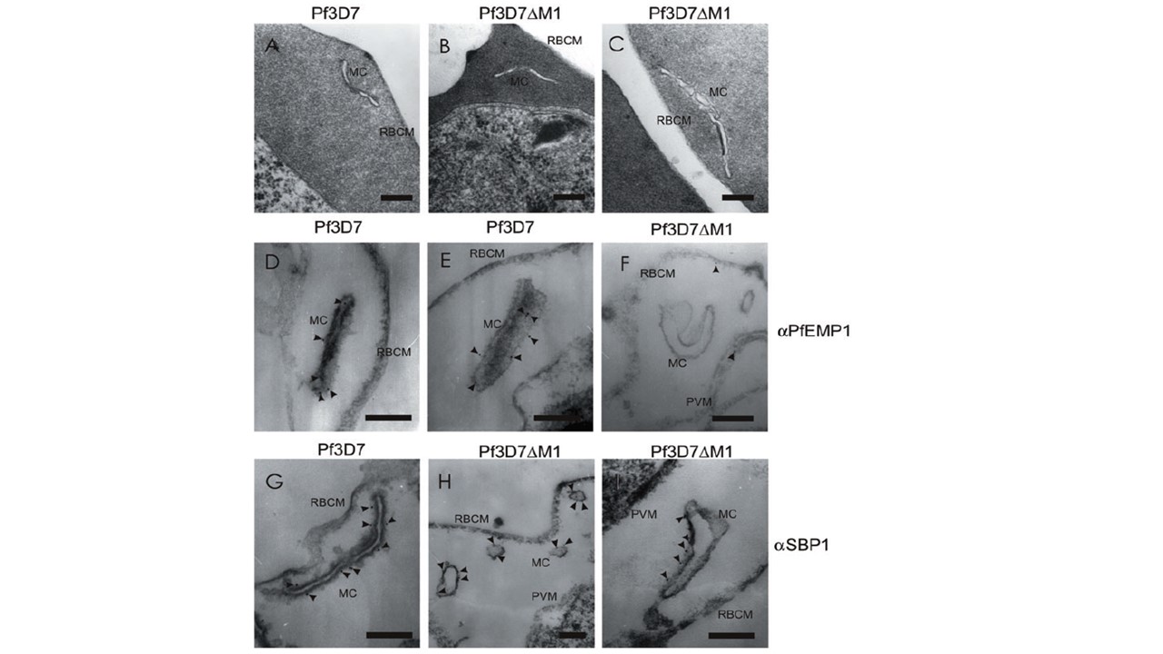
EM of 3D7 and Pf3D7DM1-infected RBCs. A–C. Sections through RBCs containing mid-trophozoite stage wild-type 3D7 parasite (A) or Pf3D7DM1 transfectant (B and C), showing Maurer’s clefts (MC) underlying the RBC membrane (RBCM). D–F. Paraformaldehyde-fixed EqtII-permeabilized RBCs infected with parent (D and E) or Pf3D7DM1 (F) parasites were labelled with anti-PfEMP1. In 3D7-infected RBCs, the Maurer’s clefts appear as long slender cisternae and the gold particles are associated with these structures. The MAHRP1-disrupted parasites are not labelled with anti-PfEMP1 although occasional gold particles are observed associated with the PVM and the cytoplasmic surface of the RBC membrane (F, arrowheads). G–I. EqtII-permeabilized RBCs infected with parent (G) or Pf3D7DM1 (H and I) parasites were labelled with anti-SBP1 antiserum followed by protein A gold (6 nm conjugate). In 3D7-infected RBCs, the gold particles are associated with the electron-dense coat of the Maurer’s cleft (G, arrowheads). In Pf3D7DM1-infected RBCs (H and I), the Maurer’s clefts appear swollen and fragmented, but are still labelled with anti-SBP1 (arrowheads). Bars are 100 nm
Spycher C, Rug M, Pachlatko E, Hanssen E, Ferguson D, Cowman AF, Tilley L, Beck HP. The Maurer's cleft protein MAHRP1 is essential for trafficking of PfEMP1 to the surface of Plasmodium falciparum-infected erythrocytes. Mol Microbiol. 2008 68(5):1300-14.
Other associated proteins
| PFID | Formal Annotation |
|---|---|
| PF3D7_0501300 | skeleton-binding protein 1 |
| PF3D7_1370300 | membrane associated histidine-rich protein |