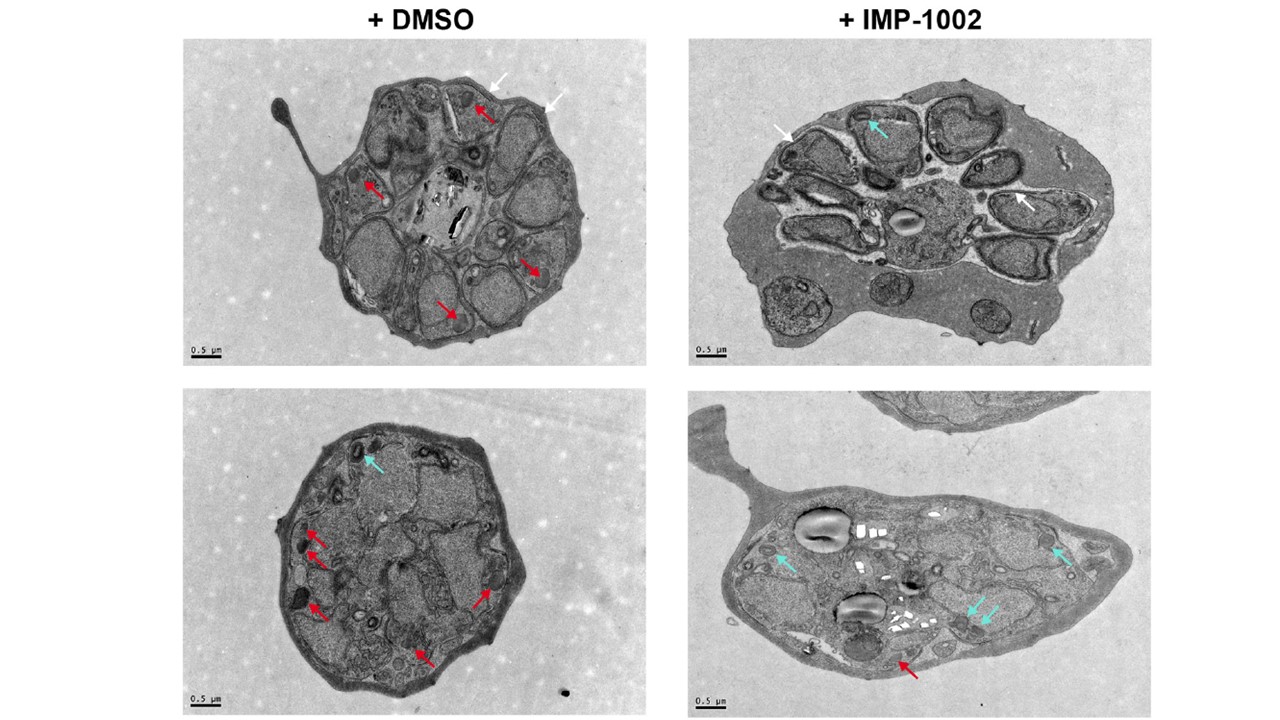
Transmission electron micrographs of DMSO and IMP-1002–treated schizonts. IMC appears to develop normally in both parasite populations (white arrows). Two populations of rhoptry were evident: electron-dense structures (red arrows) that were predominantly in control cells and those with a lighter core or inclusion (cyan arrows) found largely in drug-treated cells. Scale bar = 0.5 μm. Electron micrographs of schizonts from treated or untreated cultures revealed no gross ultrastructural changes resulting from NMT inhibition, for example, formation of the IMC occurred. However, there was a distinct difference in rhoptries. Two populations were identified, those considered “normal” and predominant in the images from the control cells and those considered as “with a lighter core/inclusion,” which were more frequent in the images of inhibitor-treated cells.
Schlott AC, Knuepfer E, Green JL, Hobson P, Borg AJ, Morales-Sanfrutos J, Perrin AJ, Maclachlan C, Collinson LM, Snijders AP, Tate EW, Holder AA. Inhibition of protein N-myristoylation blocks Plasmodium falciparum intraerythrocytic development, egress and invasion. PLoS Biol. 2021 19(10):e3001408.