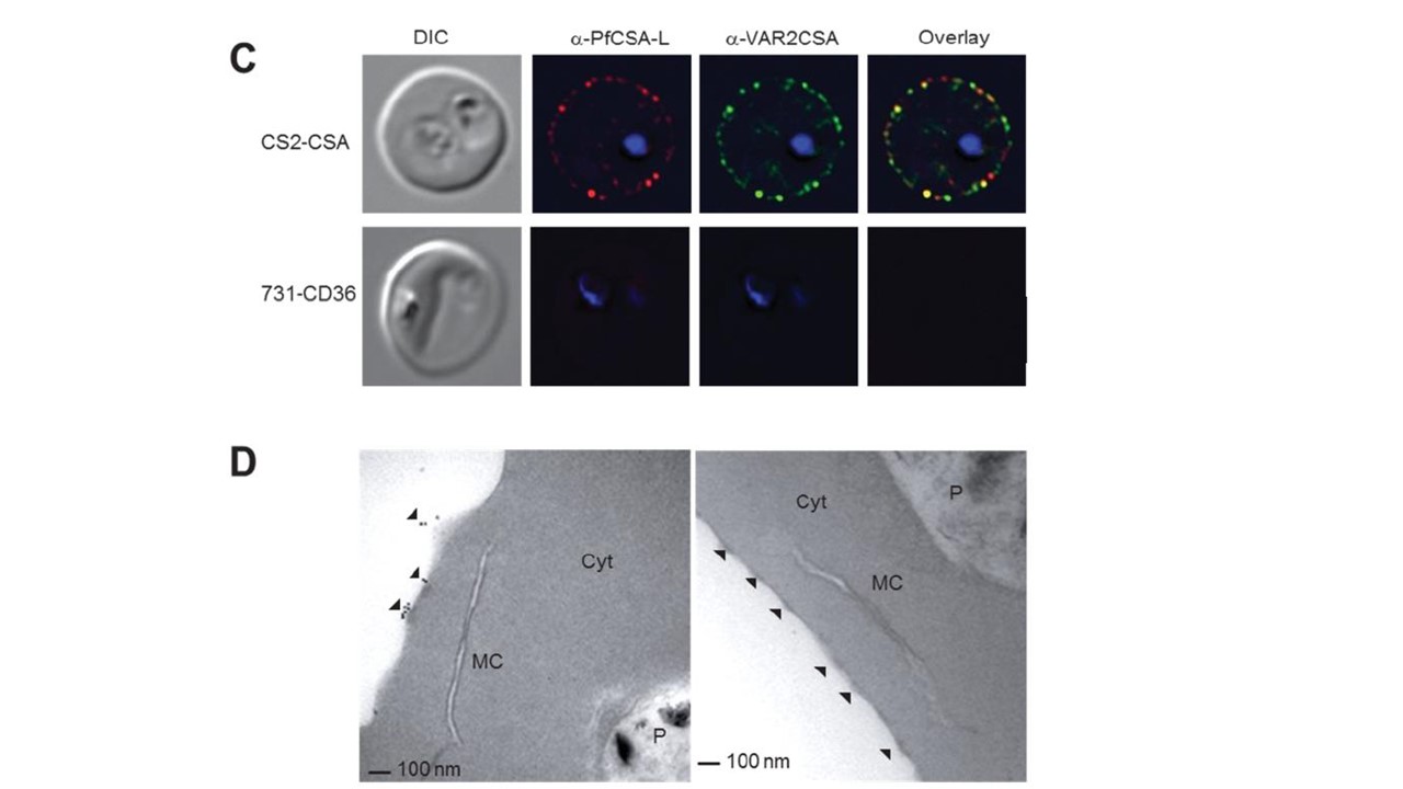
PfCSA-L is exposed on the outer surface of iRBC knobs. (C) Immunofluorescence assay (IFA) of live, intact CSA-binding iRBCs (CS2) and children’s CD36-binding iRBCs (731) using PfCSA-L monoclonal antibodies 1H11 (red) and rat α-DBL6-VAR2CSA (green) antibodies. The first and last panels represent DIC and overlay images, respectively. Yellow color indicates co-localization; blue represents DAPI nuclei staining. mAb 1H11 reacted with the surface of live intact CSA-binding, but not CD36-binding iRBCs. (D) Immunoelectron microscopy of maternal iRBC (left) and a child’s iRBC (right) after labeling with rabbit PfCSA-L-specific antisera. Black arrows point to knob-associated gold particles on iRBC surface; In agreement with the IFAs, PfCSA-L was detected on the surface knobs of CSA-binding, but not CD36-binding iRBCs (D). No reactivity was observed with control anti-AMA1 antibodiesMC, Maurer’s clefts; P, parasite; Cyt, red blood cell cytoplasm.
Keitany GJ, Jenkins BJ, Obiakor HT, Daniel S, Muehlenbachs A, Semblat JP, Gamain B, Doritchamou JYA, Desai SA, MacDonald NJ, Narum DL, Morrison R, Saveria T, Vignali M, Oleinikov AV, Fried M, Duffy PE. An invariant protein that co-localizes with VAR2CSA on Plasmodium falciparum-infected red cells binds to chondroitin sulfate A. J Infect Dis. 2021 Oct 29:jiab550
Other associated proteins
| PFID | Formal Annotation |
|---|---|
| PF3D7_1200600 | erythrocyte membrane protein 1, PfEMP1 |