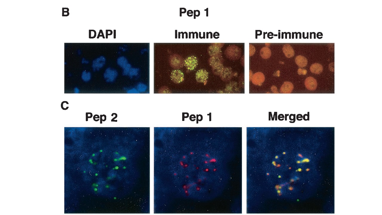
Expression of STEVOR in 3D7 parasites. (B) IFA of fixed 3D7 pRBCs (late trophozoites and schizonts). pRBCs are stained blue with DAPI nucleic acid stain (left panel) and green with immune serum (middle panel) and are not stained with preimmune rabbit polyclonal serum against peptide 1 (right panel). Following incubation with FITCconjugated goat anti-rabbit IgG, the cells were visualized with a fluorescence microscope. The left and middle panels are the same microscopic field. (C) Colocalization studies of anti-STEVOR antibodies. The images show IFA of fixed 3D7 schizonts stained with anti-peptide 2 antibodies and FITC-conjugated goat anti-mouse IgG (green) (left panel) and with anti-peptide 1 antibodies and tetramethyl rhodamine isothiocyanate conjugated goat anti-rabbit IgG (red) (middle panel). The right panel shows a merging of the two images. An Olympus Delta Vision imaging system was used (magnification, X1,000).
Kaviratne M, Khan SM, Jarra W, Preiser PR. Small variant STEVOR antigen is uniquely located within Maurer's clefts in Plasmodium falciparum-infected red blood cells. Eukaryot Cell. 2002 Dec;1(6):926-35.