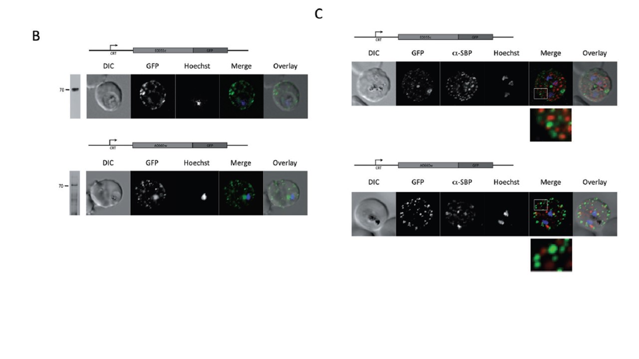
Live cell imaging and immunofluorescence microscopy of GFP transfectant lines. A, B and D. Epifluorescence microscopy of erythrocytes infected with PFE55RESA (A, left) or PFA660RESA (A, right), PFE55END (B, left) or PFA660END (B, right), PFE55INT (D, left) or PFA660INT (D, right). Both PFE55/PFA660–GFP are transported to the erythrocyte cytosol, where the chimeric proteins can be seen in ‘dot-like’ structures distributed within the host cell (B). Additionally, erythrocytes infected with PFA660CRT show a low level of background fluorescence in the erythrocyte, not associated with these punctate structures (B, lower panel).
Külzer S, Rug M, Brinkmann K, Cannon P, Cowman A, Lingelbach K, Blatch GL, Maier AG, Przyborski JM. Parasite-encoded Hsp40 proteins define novel mobile structures in the cytosol of the P. falciparum-infected erythrocyte. Cell Microbiol. 2010 12(10):1398-420.
Other associated proteins
| PFID | Formal Annotation |
|---|---|
| PF3D7_0113700 | heat shock protein 40, type II |