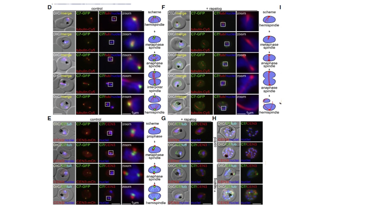
(D and F) Live cell fluorescence microscopy images (representative images of 3 independent microscopy sessions with at least 10 image series per session and condition) of synchronized (0–2 h synchronization window) Chr3C7 parasites grown in absence (D, control) and presence (F, + rapalog, induced Chr3C7 KS using the lyn mislocalizer) of rapalog and co-stained with Tubulin Tracker Deep Red during first nuclear division. Typical spindle types and duplication of Chr3C7 focus(D) and elongated anaphase spindles and incomplete nuclear segregation (F) were observed (zoom and indicated in the schemes). See Figure S4B for full panels.(E and G) Live cell fluorescence microscopy images (representative images of 3 independent microscopy sessions with at least 10 image series per session and condition) showing co-localization with centriolar plaque marker CEN3-mCherry (CEN3-mCh) in synchronized (0–2 h synchronization window) Chr3C7 knock in parasites simultaneously expressing CEN3-mCherry for co-localization and the lyn mislocalizer without fluorescent tag for KS of Chr3C7 to PPM grown in absence (E, control) and presence of rapalog (G, + rapalog, to induce Chr3C7 KS with the lyn mislocalizer) and co-stained with Tubulin Tracker Deep Red during first nuclear division. CEN3 focus duplication occurred before Chr3C7 focus duplication
Wichers-Misterek JS, Cronshagen J, Sabitzki R, Mesén-Ramírez P, Behrens HM, Bártfai R, Spielmann T. Gene-by-gene screen of the unknown proteins encoded on Plasmodium falciparum chromosome 3. Cell Syst. 2023 Jan 18;14(1):9-23.e7. doi: 10.1016/j.cels.2022.12.001. PMID: 36657393.