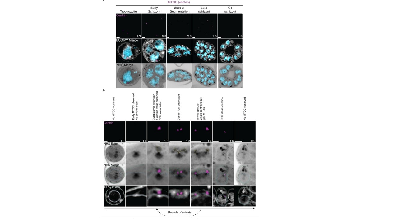
Microtubule organizing center (MTOC) biogenesis and dynamics.
(a) 3D7 parasites were prepared by U-ExM, stained with NHS ester (greyscale), BODIPY TRc (white), SYTOX (cyan) and anti-centrin (MTOC; magenta) antibodies and imaged using Airyscan microscopy across the asexual blood stage. (b) Proposed timeline of events in MTOC biogenesis, dynamics, and disassembly. Yellow line = cytoplasmic extensions, blue line = nuclear envelope,
green line = parasite plasma membrane. Images are maximum-intensity projections, number on image = Z-axis thickness of projection in µm. Scale bars = 2 µm.
Anaguano D, Frölich S, Muralidharan V, Wilson DW, Dvorin J, Absalon S. Atlas of Plasmodium falciparum intraerythrocytic development using expansion microscopy. bioRxiv [Preprint]. 2023 24:2023.03.22.533773. PMID: 36993606;