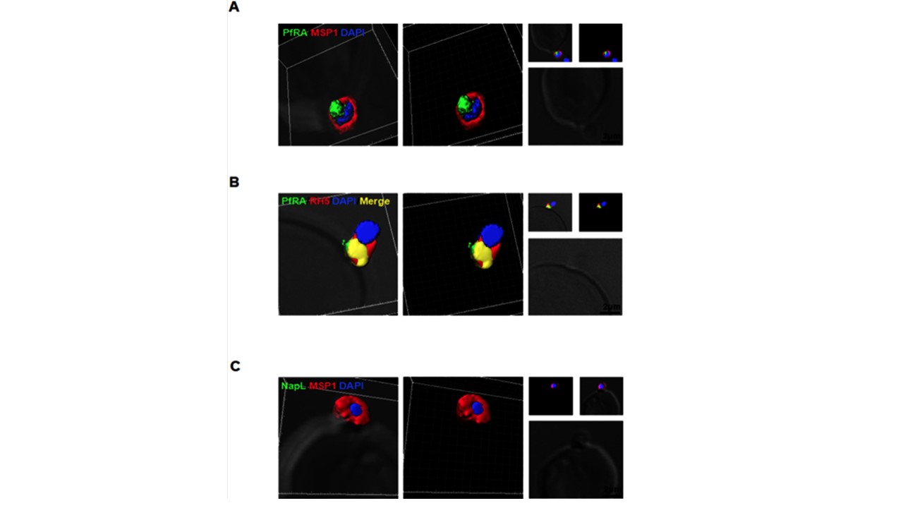
PfRA is present at the apical surface of an invading merozoite. Confocal immunofluorescence microscopy analysis of PfRA localization in cytochalasin-D treated invading merozoites under non-permeable fixing conditions using glutaraldehyde /paraformaldehyde. (A) 3D reconstruction of z-stacks during merozoite invasion co-immunostained with MSP-1 and PfRA. PfRA detected at the apical end of the merozoite surface. In the 3D images, the γ settings were altered for visual representation only. (B) PfRA (green) co-localizes with
rhoptry bulb marker PfRH5 (red) that is known to be translocated to the merozoite surface to engage with its erythrocyte receptor, Basigin. (C) As a control, staining of a nuclear parasite protein, NapL, was not detected under the same non-permeable fixing conditions confirming that the fluorescent signals were obtained only from surface localized proteins. Nuclei were stained with DNA intercalating dye DAPI (Scale bar, 2μm.)
Anand G, Reddy KS, Pandey AK, Mian SY, Singh H, Mittal SA, Amlabu E, Bassat Q, Mayor A, Chauhan VS, Gaur D. A novel Plasmodium falciparum rhoptry associated adhesin mediates erythrocyte invasion through the sialic-acid dependent pathway. Sci Rep. 2016 6:29185. PMID: 27383149;
Other associated proteins
| PFID | Formal Annotation |
|---|---|
| PF3D7_0424100 | reticulocyte binding protein homologue 5 |
| PF3D7_0930300 | merozoite surface protein 1 |
| PF3D7_1203700 | nucleosome assembly protein |