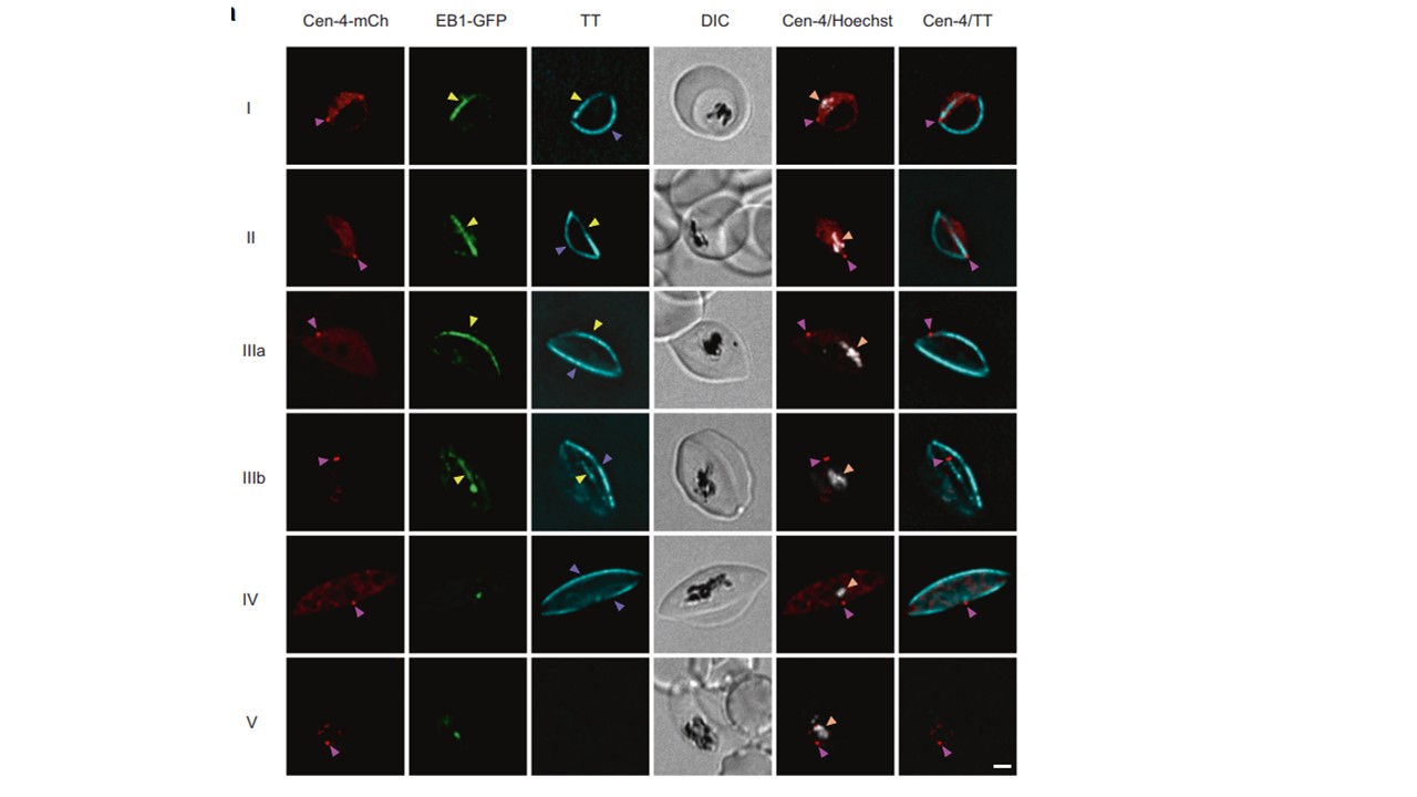
The gametocyte MTOC is a centriolar plaque equivalent. a Live-cell
fluorescence imaging of stage I-V gametocytes in the PfEB1-GFP/Pfcentrin-4-mCherry co-transfectant parasite line. Pfcentrin-4-mCherry (Cen-4-mCh, red, magenta arrows) delineates the punctate centriolar plaque and PfEB1-GFPdecorated (EB1-GFP, green)/Tubulin Tracker (TT, cyan)-labelled nuclear microtubules (yellow arrowheads) appear to emanate from this structure in early stage gametocytes (stage I, II). As the gametocyte develops, the sub-pellicular microtubule population (purple arrowheads) appears to move away from the Pfcentrin-4-mCherry punctum (stage III). In stage IV, the sub-pellicular microtubules form a cage around the gametocyte while the nuclear microtubules collapse. Hoechst (grayscale, orange arrowheads) is used to label chromatin. Differential interference contrast (DIC) images are shown. Scale bars: 2 μm
Li J, Shami GJ, Cho E, Liu B, Hanssen E, Dixon MWA, Tilley L. Repurposing the mitotic machinery to drive cellular elongation and chromatin reorganisation in Plasmodium falciparum gametocytes. Nat Commun. 2022 13(1):5054.
Other associated proteins
| PFID | Formal Annotation |
|---|---|
| PF3D7_0307300 | microtubule-associated protein RP/EB family, putative |