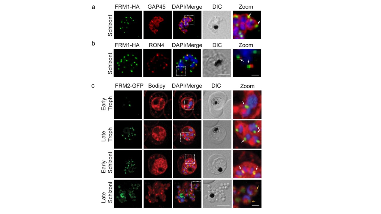
FRM1 and 2 show different cellular locations in asexual stage parasites. a Immunofluorescence microscopy of FRM1-HA parasites labelled with anti-HA (green) and anti-GAP45 (red). b anti-HA (green) and RON4 (red). DAPI (blue), merge and Differential interference contrast (DIC) images are shown. Scale bar = 5 μm. Dotted white line outlines the zoom panel. Zoom scale bar = 1 μm. c Live-cell immunofluorescence of FRM2-GFP (green) co-labelled with BODIPY-TR-Ceramide (Bodipy, red). DAPI (blue), merge and DIC images are shown. The dotted white line outlines the zoom panel. Zoom Scale bar = 1 μm.
PubMed Article: Plasmodium falciparum formins are essential for invasion and sexual stage development
Other associated proteins
| PFID | Formal Annotation |
|---|---|
| PF3D7_0530900 | formin 1 |
| PF3D7_1219000 | formin 2 |
| PF3D7_1222700 | glideosome-associated protein 45 |