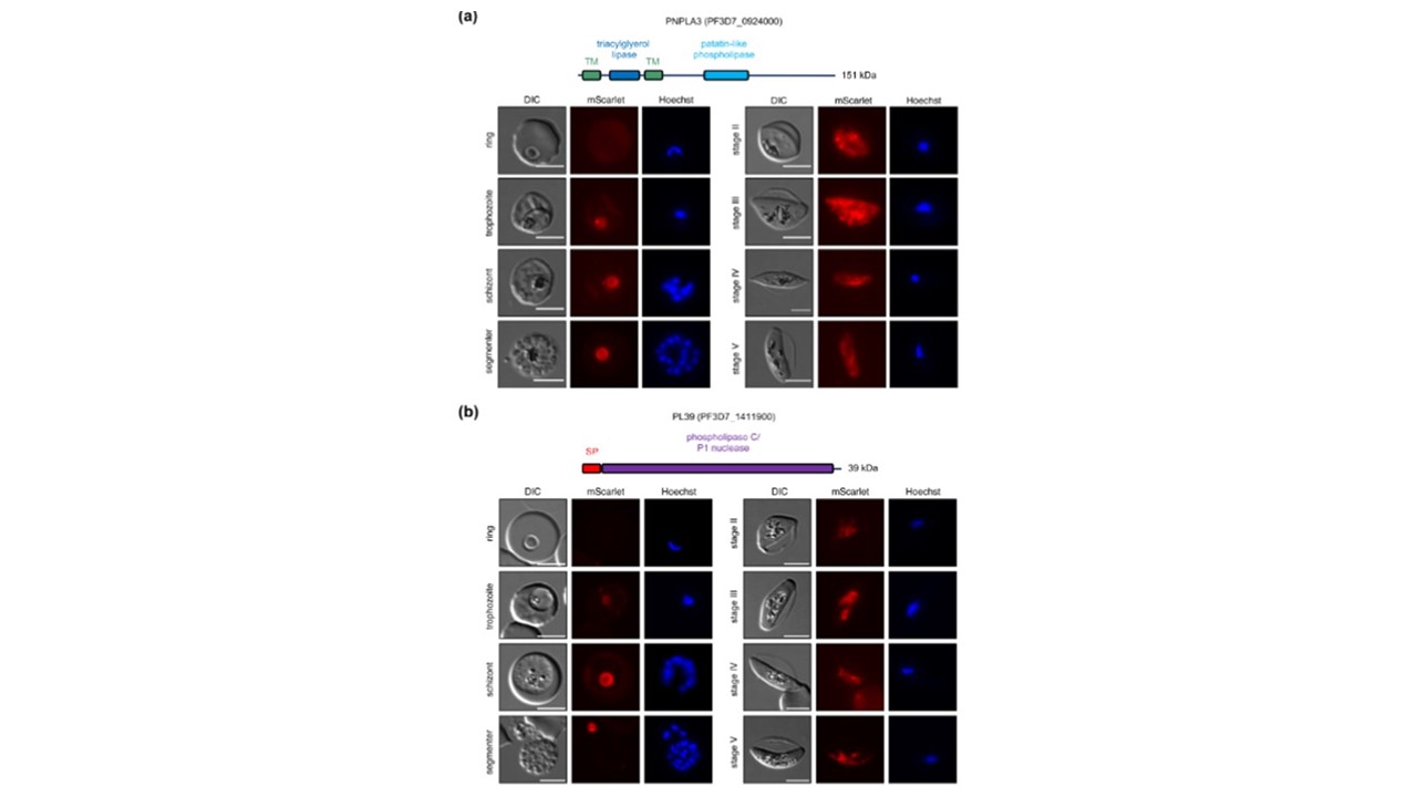
Localization analysis of PNPLA3 and PL39. Parasites expressing endogenously tagged PNPLA3-mScarlet (a) and PL39-mScarlet (b) were analyzed during asexual and sexual blood stage development by live-cell microscopy. Nuclei were stained with Hoechst. Scale bars = 5 μm. DIC, differential interference contrast. Schematic representations of the functional domains of the two proteins are shown on top of the images. SP, signal peptide; TM, transmembrane domain.
Pietsch E, Niedermüller K, Andrews M, Meyer BS, Lenz TL, Wilson DW, Gilberger TW, Burda PC. Disruption of a Plasmodium falciparum patatin-like phospholipase delays male gametocyte exflagellation. Mol Microbiol. 2023
PubMed Article: Disruption of a Plasmodium falciparum patatin-like phospholipase delays male gametocyte exflagellation
Other associated proteins
| PFID | Formal Annotation |
|---|---|
| PF3D7_0924000 | patatin-like phospholipase, putative |