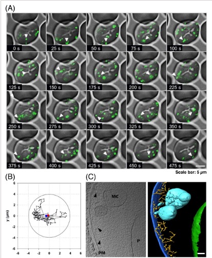
The Maurer's clefts are established during the ring stage of P. falciparum and move throughout the cytoplasm of the infected RBC via Brownian motion. (A) Movement of MC in a RBC infected with ring stages of P. falciparum. The shown parasite strain is 3D7 which expresses a genomically integrated fusion of the MC marker protein PfSBP1 (P. falciparum skeleton-binding protein 1) with GFP. The time series shows an infected RBC over a time period of 494 s (time interval between each data point: 3.5 s). Scale bar: 5 µm. (B) Total projection of the movement of the MC shown in the previous section reveals the Brownian motion of the observed organelle. The circle represents the border of the infected RBC. (C) Single image of a cyro-electron tomogram (left) with the corresponding surface-rendered view (right) of a MC in a red blood cell infected with the ring stage of P. falciparum. Labeling and color code of the surface-rendered view. PM: plasma membrane of the RBC (dark blue); MC: Maurer's cleft (cyan); P, parasite (green), actin filaments (yellow). Arrowheads show the actin filaments. Scale bar: 100 nm.
Bekić V, Kilian N. Novel secretory organelles of parasite origin - at the center of host-parasite interaction. Bioessays. 2023 Jul 30:e2200241.