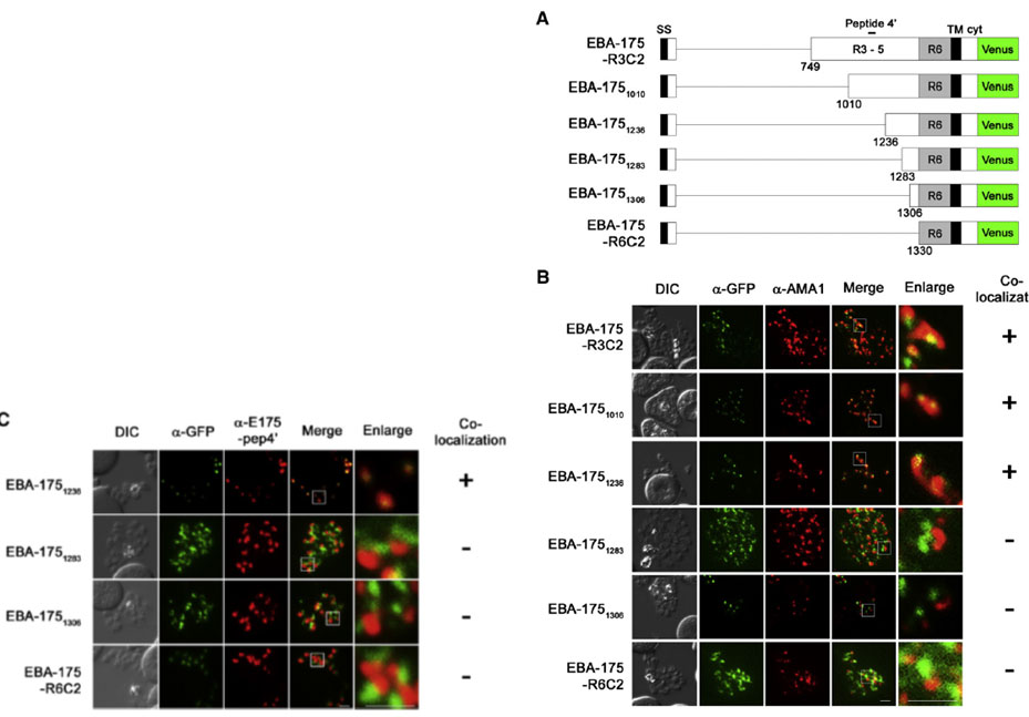
IFA detection of parasites expressing serially truncated recombinant EBA-175. (A) Schematic structures of recombinant EBA-175 proteins; EBA-175-R3C2, EBA-1751010, EBA-1751236, EBA-1751283, EBA-1751306 or EBA-175-R6C2. Signal sequence (SS), regions 1 to 6 (R1, R2, R3–5 and R6), transmembrane region (TM) and cytoplasmic tail (cyt) are indicated. Bar above the scheme indicates the target region of anti-EBA-175-pep4′ antibody (aa 1089–1108). (B and C) Parasites were stained with anti-GFP (α-GFP, green) and anti-AMA1 (panel B; α-AMA1, red) or anti-EBA-175-pep4′ (panel C; α-E175-pep4′, red). Scale bar indicates 2 μm. DIC is a differential interference contrast image. Status of the co-localization with a microneme marker is shown at the right side of each row. (+) or (−) indicate that two signal co-localized or not co-localized.
Sakura T, Yahata K, Kaneko O. The upstream sequence segment of the C-terminal cysteine-rich domain is required for microneme trafficking of Plasmodium falciparum erythrocyte binding antigen 175. Parasitol Int. 2012 62(2):157-64
Other associated proteins
| PFID | Formal Annotation |
|---|---|
| PF3D7_0731500 | erythrocyte binding antigen-175 |