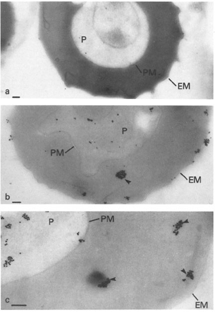
Immunoelectron microscopy of mature trophozoites of K + Malayan Camp P. falciparum with (a) a control IgM mAb (1A1D5) or (b and c) mAb 87. In a, the ascites was diluted to 1:400, and in b and c, the ascites was diluted to 1:6,000 for reaction with the frozen section. Addition of secondary antibody (rabbit anti-mouse Ig) prior to Protein A-gold had no effect on the results (a and c show the results without secondary antibody). Identical results to a were obtained if primary antibody was omitted and Protein A-gold added either directly or after incubation with secondary antibody. P, parasite; PM, parasite plasma membrane; EM, erythrocyte plasma membrane. Dense packets of Protein A-gold particles reacting with the cytoplasm of the host erythrocyte are indicated by arrows in b and c. Bars, 0.5 mm.
Pf HRP II localized to several cell compartments including the parasite cytoplasm, as concentrated "packets" in the host erythrocyte cytoplasm and at the IRBC membrane. The results provide evidence for an intracellular route of transport for a secreted malarial protein from the parasite through several membranes and the host cell cytoplasm.
Howard RJ, Uni S, Aikawa M, Aley SB, Leech JH, Lew AM, Wellems TE, Rener J, Taylor DW. Secretion of a malarial histidine-rich protein (Pf HRP II) from Plasmodium falciparum-infected erythrocytes. J Cell Biol. 1986 103:1269-77.