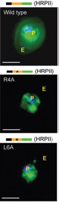
Fluorescent micrographs of live cells expressing green fluorescent protein (GFP) chimeras of the parasite vacuolar translocation sequence containing the wild-type HT motif or point mutations at R4A or L6A, where 4 and 6 indicate the position in the signal. Export of GFP to the erythrocyte (E) is abrogated by both mutations. GFP fluorescence was detected by digitized fluorescence microscopy. Parasite (P) nucleus is Hoechst stained (blue). Schematics above micrographs indicate constructs containing an endoplasmic reticulum–type signal sequence (black) needed for protein delivery to the parasitophorous vacuolar membrane, the vacuolar translocation sequence (orange) of the indicated protein, and the C-terminal GFP protein (green). Stars indicate single amino acid substitutions. Scale bar, 5 μm. HRPII, histidine-rich protein II.
Haldar K, Murphy SC, Milner DA, Taylor TE. Malaria: mechanisms of erythrocytic infection and pathological correlates of severe disease. Annu Rev Pathol. 2007; 2:217-49.