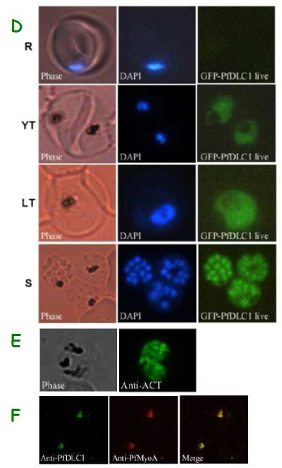
Expression of PfDLC1, PfACT1, and PfMyoA gene products by wild type and transfected P. falciparum. D. Expression and localization of PfDLC1-GFP throughout the erythrocytic cell cycle of P. falciparum. Live transfected parasites were analysed by fluorescence microscopy. E. Immunofluorescence localization of P. falciparum actin in fixed transgenic parasites (late trophozoites). F. Co-localization of PfDLC1 and PfMyoA. Smears of erythrocytes infected with 3D7 strain were fixed and probed with rat anti-PfDLC1 followed by anti-rat IgG-FITC and rabbit anti-PfMyoA, followed by anti-rabbit IgG-Alexa Fluor 568 (red). PfDLC1 location showed a homogenous distribution in the cytoplasm of the parasite (D). It seems that the protein is not exported to the nucleus of P. falciparum or to the RBC cytoplasm or to the RBC surface (Fig. 7C and 7D). The expression of PfDLC1 in younger stage transfectant as well as in the wild type stage (young ring) was undetectable (D). P. falciparum actin is distributed in the parasite cytoplasm (E). Myosin A partially overlap PfDLC1 in schizonts (F).
Daher W, Pierrot C, Kalamou H, Pinder JC, Margos G, Dive D, Franke-Fayard B, Janse CJ, Khalife J. Plasmodium falciparum dynein light chain 1 interacts with actin/myosin during blood stage development. J Biol Chem. 2010 285:20180-20191.
Other associated proteins
| PFID | Formal Annotation |
|---|---|
| PF3D7_0822500 | leucine-rich repeat protein dynein light chain 1 |
| PF3D7_1342600 | myosin A |