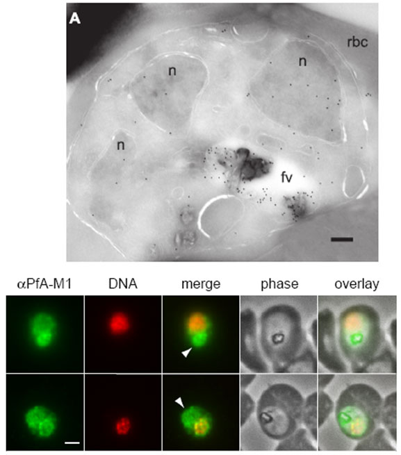
Upper: Immunofluorescence localization of PfA-M1 in parasites fixed with 50% methanol/50% ethanol at -20 °C. Anti-PfA-M1 fluorescence is pseudocolored green and DAPI fluorescence (DNA) is pseudocolored red. Food vacuole fluorescence is identified by co-localization with the hemozoin crystal and is indicated with an arrowhead in the “merge” image. Panel B clearly shows PfA-M1 label in the food vacuole, whereas a nuclear localization is more evident in panel C. Scale bar, 1 μm.
Lower: Localization of PfA-M1 in intraerythrocytic P. falciparum. A, PfA-M1 was detected in a parasite cryosection with affinity-purified anti-PfA-M1 antibodies. PfA-M1 is present throughout the lumen of the food vacuole and in the nucleus. fv, food vacuole; n, nucleus; rbc, red blood cell. Scale bar, 250 nm
Ragheb D, Dalal S, Bompiani KM, Ray WK, Klemba M. Distribution and Biochemical Properties of an M1-family aminopeptidase in Plasmodium falciparum indicate a role in vacuolar hemoglobin catabolism. J Biol Chem. 2011 286:27255-65