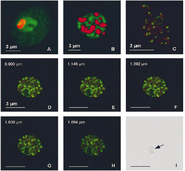
PfA-M1 subcellular location. Samples from Plasmodium falciparum in vitro cultures at various stages were fixed in a mix of methanol and acetone (1:4) and the fuorescence associated with the anti-MAP1 Igs (10 mg/ml, green) and with the nuclei (red) was observed by confocal microscopy. (A) Trophozoite. (B) Schizont. (C) Released merozoites. (D-H) Series of 5 optical sections through a mature schizont (positions along the z axis indicated on the upper left of each panel). (I) Corresponding phase-contrast image, showing the digestive vacuole (indicated by an arrow). In trophozoites, the labelling was diffuse in the parasite cytoplasm, with accumulations around the food vacuole. In schizonts, it turned progressively to a vesicle-like pattern, ending as a clear spot in released merozoites.
Allary M, Schrevel J, Florent I. Properties, stage-dependent expression and localization of Plasmodium falciparum M1 family zinc-aminopeptidase. Parasitology. 2002 125:1-10. Copyright Cambridge University Press.