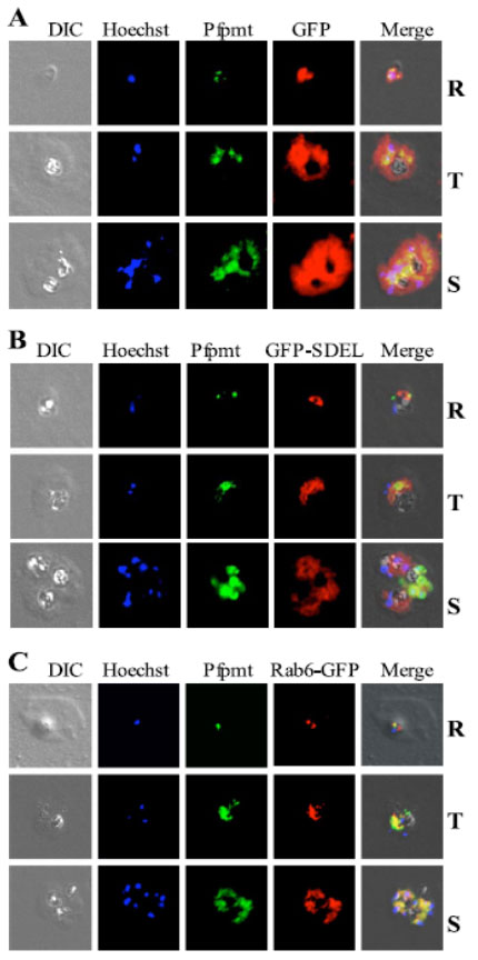
Immunofluorescence microscopy of Pfpmt in transgenic P. falciparum-infected red blood cells expressing GFP in the cytoplasm, ER, and Golgi apparatus. Erythrocytes infected with transgenic parasites expressing GFP in the cytoplasm (GFP) ( panel A), the ER (GFP-SDEL-PF14_0046) (panel B), and Golgi apparatus (Rab6-GFP PF11_0461) (panel C) at different stages of parasite intraerythrocytic development. Parasites were examined by microscopy using illumination at 546 nm to visualize Pfpmt (green) conjugated to the fluorescein isothiocyanate-conjugated anti-rabbit secondary antibody or at 488 nm to visualize GFP complexed with the Texas Red-conjugated anti-mouse secondary antibody (red). DNA was counterstained with Hoechst (blue). DIC, differential interference contrast images of parasitized erythrocytes. R, ring; T, trophozoite; S, schizont.
Confocal microscopy revealed that Pfpmt is not cytoplasmic and complete co-localization was detected with Rab6, a marker of the Golgi apparatus.
Witola WH, Pessi G, El Bissati K, Reynolds JM, Mamoun CB. Localization of the phosphoethanolamine methyltransferase of the human malaria parasite Plasmodium falciparum to the Golgi apparatus. J Biol Chem. 2006 281:21305-11.
Other associated proteins
| PFID | Formal Annotation |
|---|---|
| PF3D7_1343000 | phosphoethanolamine N-methyltransferase |
| PF3D7_1404900 | conserved Plasmodium protein, unknown function |