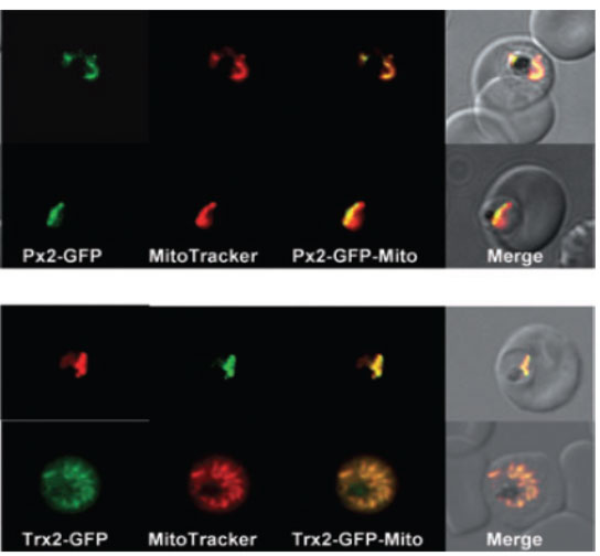
Localization of PfTrx-Px2 and PfTrx2 in P. falciparum erythrocytic stages. P. falciparum erythrocytic stages were transfected with construct pHH2-Px2-GFP, pHH2-Trx2-GFP leading to the expression of the peroxiredoxin or thioredoxin2 C-terminally fused to green fluorescent protein (GFP).
A. The localization of the peroxiredoxin-GFP fusion protein in parasites previously treated with MitoTracker CMX-Ros was analysed by fluorescence light microscopy. Phase, phase contrast of parasitized erythrocytes infected with P. falciparum trophozoites; PfTrx-Px2-GFP, parasitized erythrocytes expressing the peroxiredoxin-GFP fusion protein analysed using the FITC channel; MitoTracker, parasitized erythrocytes expressing the peroxiredoxin-GFP fusion protein analysed using the rhodamine channel; PfTrx-Px2-Mito; merge of FITC and rhodamine channels; merge, merge of all images. The images show that the peroxiredoxin-GFP fusion protein is colocalizing with the mitochondrion (stained by MitoTracker).
B. The expression of pHH2-PfTrx2 results in the localization of the fusion protein the mitochondrion. Phase, phase contrast of parasitized erythrocytes infected with P. falciparum trophozoites; PfTrx2-GFP, parasitized erythrocytes expressing the Trx2-GFP fusion protein analysed using the FITC channel; MitoTracker, parasitized erythrocytes expressing the Trx2-GFP fusion protein analysed using the rhodamine channel; PfTrx2-Mito; merge of FITC and rhodamine channels; merge, merge of all images.
Boucher IW, McMillan PJ, Gabrielsen M, Akerman SE, Brannigan JA, Schnick C, Brzozowski AM, Wilkinson AJ, Müller S. Structural and biochemical characterization of a mitochondrial peroxiredoxin from Plasmodium falciparum. Mol Microbiol. 2006 61:948-59.PMID
Other associated proteins
| PFID | Formal Annotation |
|---|---|
| PF3D7_1215000 | thioredoxin peroxidase 2 |