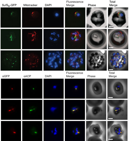
Subcellular localization of SufS to the apicoplast. Upper panel) Epifluorescent images of live P. falciparum erythrocytic-stage parasites expressing GFP fused to the SufS leader peptide (SufSlp-GFP). The parasites were stained with mitotracker to identify mitochondria and DAPI to identify nuclei. Image z-stacks were deconvolved and then presented as a single combined image. Scale bar = 2 mm. GFP fluorescence localizes to an elongated organelle distinct from the mitochondrion in late ring (top row), late trophozoite or early schizont (middle), and schizont (bottom) stage parasites. Lower panel) Immunofluorescence co-localization of SufSlp-GFP and endogenous ACP. An antibody specific for GFP co-localized with αACP antibodies, demonstrating apicoplast localization in late ring (top panel), late trophozoite or early schizont (middle), and schizont (bottom) stage parasites. The parasites were stained with DAPI to identify nuclei. Image z-stacks were deconvolved and then presented as a single combined image. Scale bar = 2 µm.
Gisselberg JE, Dellibovi-Ragheb TA, Matthews KA, Bosch G, Prigge ST. The Suf Iron-Sulfur Cluster Synthesis Pathway Is Required for Apicoplast Maintenance in Malaria Parasites. PLoS Pathog. 2013 9(9):e1003655
Other associated proteins
| PFID | Formal Annotation |
|---|---|
| PF3D7_0208500 | acyl carrier protein |