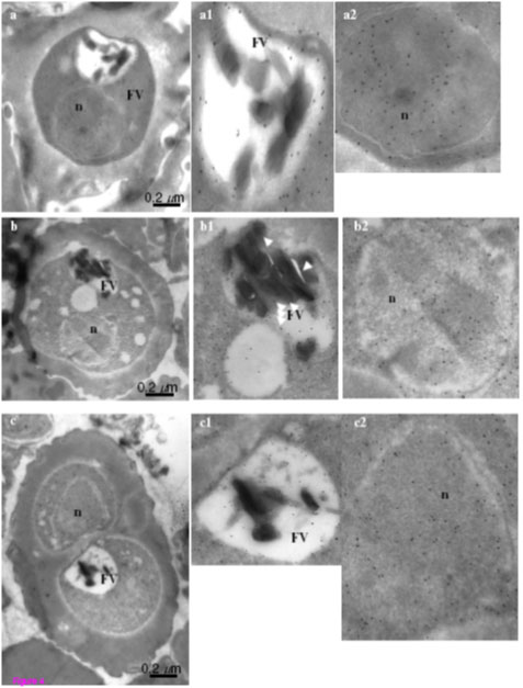
Figure 4. Immuno-gold electron microscopic (IEM) imaging for the localization of enolase in early trophozoite satge of P. falciparum using mouse anti-r-Pfen antibody. Magnified views of the food vacuole (FV) and nucleus (n) are also shown. Arrows in food vacuole marks hemozoin associated enolase. Enolase was found to be associated with cytosol,nucleus, food vacuole, cytoskeleton and plasma membrane.
Pal Bhowmick I, Kumar N, Sharma S, Coppens I, Jarori GK. Plasmodium falciparum enolase: stage-specific expression and sub-cellular localization. Malar J. 2009 8(1):179. PubMed