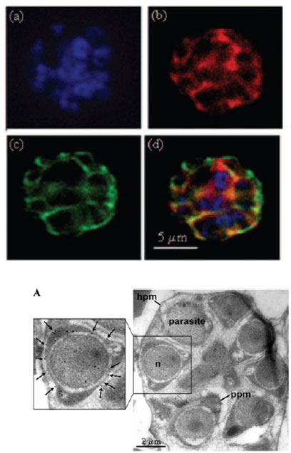
IFA of the schizont stage of P. falciparum using DAPI (a), rabbit anti-r-Pfen antiserum (1:200) with secondary anti-rabbit IgG conjugated with AlexaFluor 568 (b), and mouse anti-MSP-1 antibodies (1:100) with secondary anti-mouse IgG conjugated with AlexaFluor 488 (c). An overlay of the images from frames a, b, and c is shown in frame d. The enolase protein is localized on the merozoite cell surface.
A. IEM image using mouse anti-r-Pfen antiserum at a 1:100 dilution with a P. falciparum-infected red cell at young trophozoite stage the presence of enolase on the parasite plasma membrane (ppm) is marked with arrows. Hpm, host cell plasma membrane; n, nucleus.
Pal-Bhowmick I, Mehta M, Coppens I, Sharma S, Jarori GK. Protective properties and surface localization of Plasmodium falciparum enolase. Infect Immun. 2007 75:5500-8.