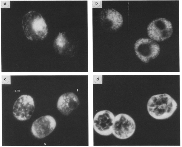
Immunofluorescent localization of GBP-130 at different stages. Thin smears of cultures overlaid with R311 serum and FITC-goat anti-rabbit serum. (a, c) cells fixed with acetone; (b, d) cells fixed with 1% paraformaldehyde; (a, b) trophozoite stage parasites; (d) segmenter stage parasites; (c) three different stages: schizont (s), segmenter (sm), trophozoite (t). Magnification: X 950. Differences in antigen localization are apparent depending on the methods used to fix to the parasites. In parasites fixed in acetone (Figs. a, c) the distribution of the antigen as detected by fluorescence is more diffuse. Immunofluorescence patterns shown in Figs. b and d are of cells fixed with paraformaldehyde. More internal structure is apparent in parasites fixed in this manner. At the trophozoite stage fixed in acetone (Fig. a) the antigen appears to be localized on the parasite and in small inclusions in the erythrocyte cytoplasm. In late trophozoites fixed in paraformaldehyde (Fig. b) the fluorescent staining is more intense and appears predominantly in vesicles or inclusions in the erythrocyte cytoplasm and perhaps the parasitophorous vacuole. The antigen is not present in uninfected erythrocytes. In mature schizonts (segmenters) fixed in paraformaldehyde (Fig. d) the antigen is localized principally in the space between the schizont and the erythrocyte membrane but also appears around the periphery of the merozoite.
Perkins M. Stage-dependent processing and localization of a Plasmodium falciparum protein of 130,000 molecular weight. Exp Parasitol. 1988 65:61-8. Copyright Elsevier 2010.