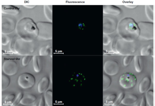
Redistribution of PfATG8 protein (green spots) upon starvation-induced cell death. The top three panels are strain HB3 Plasmodium falciparum grown in red blood cell culture under normal conditions; the bottom is a parasite from the same culture after being placed in starvation media (no sugar, no amino acids) for 6 h. The primary antibody was rabbit anti-TgATG8; the secondary was goat antirabbit IgG conjugated to DyLight488 fluorophore. Parasite nuclei were also stained with DAPI (blue) and mounted using Fluorogel mounting media. Samples were imaged using a spinning disk confocal microscope and 405-nm and 491-nm laser lines at 200 ms exposure and 35% laser power. Images were iteratively deconvoluted using an experimental point spread function and AutoQuantX2 software and displayed using Imaris 7.4.2. Controls (titrating antibodies, no primary antibody vs no secondary antibody) reveal staining is specific for PfATG8.
Sinai AP, Roepe PD. Autophagy in Apicomplexa: a life sustaining death mechanism? Trends Parasitol. 2012 28(9):358-64.