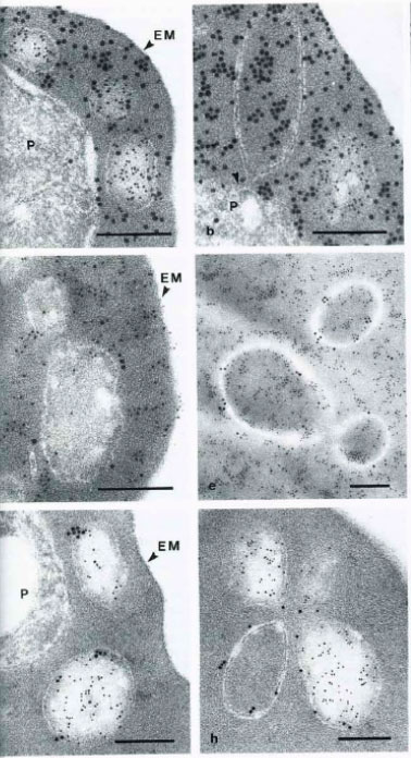
Immunoelectron micrographs of P. falciparum-infected erythrocytes showing double labeling results using anti-parasite and anti-host cell antibodies reacted with colloidal gold IgG of different particle sizes. A-B Abs directed against S-antigen (10 nm) and erythrocyte material (15 nm); D-E Abs against Exp-1 (15 nm) abd erythrocyte material (10 nm); G-H Abs against Exp-1 (15 nm) and against S-antigen (10 nm). EM – erythrocyte membrane; P – parasite; Arrowheads indicate pvm. Multiple species of vesicles, each with specifically packaged contents, are consistent with a sorting function of vesicular structures in the Plasmodium infected erythrocyte. During schizogony, two parasite antigens, an S-antigen and a parasitophorous vacuole membrane antigen, EXP-1, become packaged into such vesicles and are transported into the erythrocyte cytoplasm. At this stage of parasite development, host cell material is taken in through the parasitophorous vacuole membrane into the vacuolar space surrounding the parasite.
Stenzel DJ, Kara UA. Sorting of malarial antigens into vesicular compartments within the host cell cytoplasm as demonstrated by immunoelectron microscopy. Eur J Cell Biol. 1989 49:311-8. Copyright Elsevier 2010.
Other associated proteins
| PFID | Formal Annotation |
|---|---|
| PF3D7_1035200 | S-antigen |