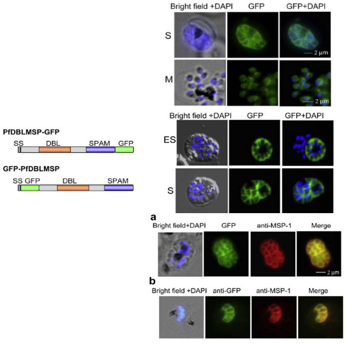
(D, E) Sub-cellular distribution of of PfDBLMSP on the merozoite surface. Fluorescent microscopic images of live parasites expressing PfDBLMSPGFP (D) and GFP-PfDBLMSP (E) fusion proteins. Both the fusion proteins (green) are localized on the surface of the merozoites. The nucleus is stained with DAPI (blue). ES, early schizont; S, schizont; and M, Merozoites. (F) Co-localization studies of GFP fusion proteins with the surface marker MSP-1 PFI1475w by immunofluorescence. The PfDBLMSP-GFP (a) and GFP-PfDBLMSP (b) fusion proteins (green) co-localize with the surface protein MSP-1 (red) as shown in the merge picture. Nuclei are stained with DAPI (blue).
Schematic domain structure of PfDBLMSP as N- or C-terminal GFP fusion, labeled as PfDBLMSP-GFP and GFP-PfDBLMSP, respectively. Signal peptides: black; green fluorescent protein: green; Duffy binding like (DBL) domain: brown; secreted polymorphic antigen associated with merozoites (SPAM) domain: blue.
Wickramarachchi T, Cabrera AL, Sinha D, Dhawan S, Chandran T, Devi YS, Kono M, Spielmann T, Gilberger TW, Chauhan VS, Mohmmed A. A novel Plasmodium falciparum erythrocyte binding protein associated with the merozoite surface, PfDBLMSP. Int J Parasitol. 2009 39:763-73. Copyright Elsevier 2009.