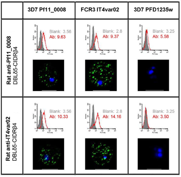
Antibody cross-reactivity to DC5-PfEMP1-expressing 3D7 and FCR3, shown using flow cytometry and confocal microscopy. Flow cytometry histograms showing the reactivity of parasite lines expressing 3D7 PF11_0008 (3D7-DC5), FCR3 IT4var02 (FCR3-DC5) and 3D7 PFD1235w (3D7-DC4) with antibodies against domains DBLδ5-CIDRβ4 from 3D7-DC5 or FCR3-DC5. Below the flow cytometry histograms are confocal microscopy pictures of the same infected erythrocytes. Surface reactivity with FITC-labeled rat antibodies is seen as green dots and the DNA in the nuclei is stained blue by DAPI. Flow cytometry assays with live parasites showed that most of the parasites in these lines expressed the native DC5-containing PfEMP1 on the surface of the infected erythrocytes, and confocal microscopy confirmed that the staining pattern was similar to that previously published for PfEMP1 Interestingly, IgG against the DBLδ5-CIDRβ4 proteins were cross-reactive since the antibody reagents stained native DC5-PfEMP1 on the surface of erythrocytes infected with both the FCR3-DC5 and the 3D7-DC5 parasite line.
Berger SS, Turner L, Wang CW, Petersen JE, Kraft M, Lusingu JP, Mmbando B, Marquard AM, Bengtsson DB, Hviid L, Nielsen MA, Theander TG, Lavstsen T. Plasmodium falciparum expressing domain cassette 5 type PfEMP1 (DC5-PfEMP1) bind PECAM1. PLoS One. 2013 8(7):e69117.
Other associated proteins
| PFID | Formal Annotation |
|---|---|
| PF3D7_0425800 | erythrocyte membrane protein 1, PfEMP1 |
| PfEMP1 | PfEMP1 |