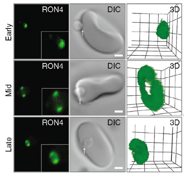
Confocal microscopy of early, mid and late merozoite invasion, showing (left ) single slice immunofluorescence of the tight junction labeled with anti-RON4, (middle) accompanying DIC images of the same invasion event and (right) a 3D projection of the anti-RON4 tight junction labeling. Arrows mark the relative position of tight junction labeling. Scale bar = 1 mm. Inset shows 2× zoom of merozoite. In 3D projections, grid = 0.5 mm and gamma settings have been changed for display purposes.
Riglar DT, Baum J. Static and Dynamic Imaging of Erythrocyte Invasion and Early Intra-erythrocytic Development in Plasmodium falciparum. Methods Mol Biol. 2013;923:269-80.