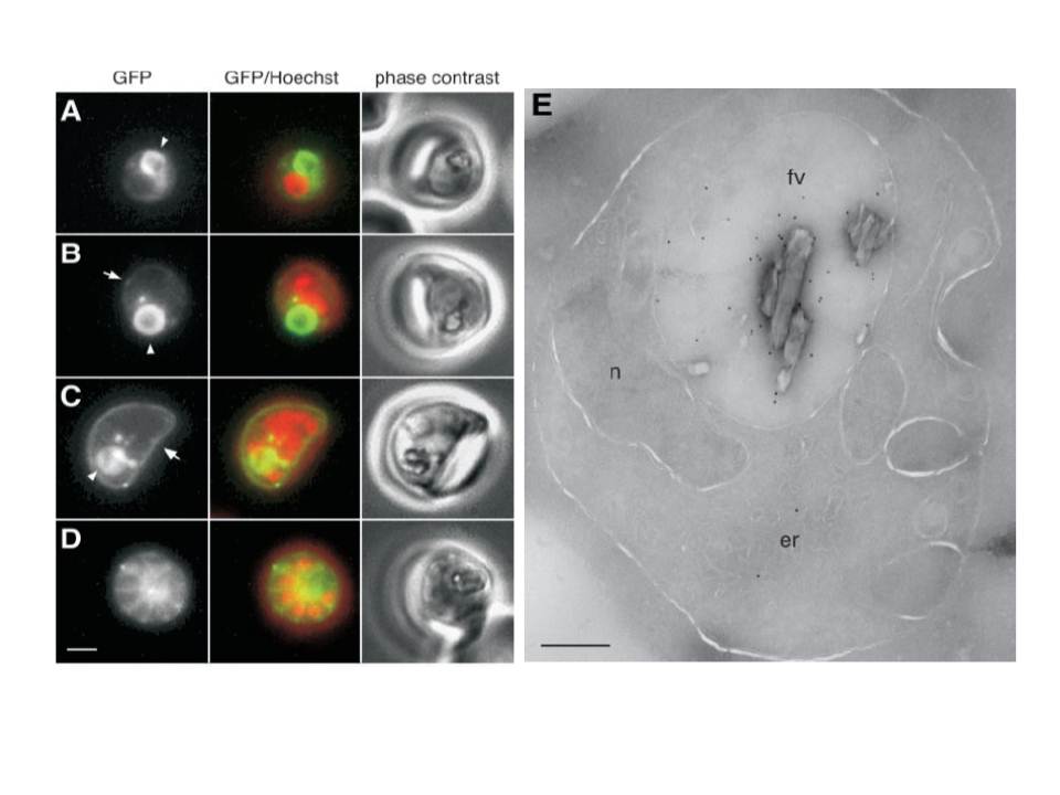
Localization of DPAP1 to the food vacuole and the parasitophorous vacuole. A–D, fluorescence microscopy of live parasites expressing DPAP1-GFP. A, trophozoite. B, ~2N schizont. C, ~8N schizont. D, 8N segmented schizont. In the center column fluorescence from GFP is green and that from nuclear stain Hoechst 33342 is red. The food vacuole is indicated with arrowheads in the GFP panels of A–C, and the parasitophorous vacuole is indicated with arrows in B and C. Fluorescent spots in B and C may be vesicular structures involved in transport of DPAP1-GFP to the food vacuole. Bar, 2 mm. E, immunogold labeling of an F9 parasite with anti-DPAP1 antibody. fv, food vacuole; er, endoplasmic reticulum; n, nucleus. Bar, 400 nm.
Klemba M, Gluzman I, Goldberg DE. A Plasmodium falciparum dipeptidyl aminopeptidase I participates in vacuolar hemoglobin degradation. J Biol Chem. 2004 279:43000-7.