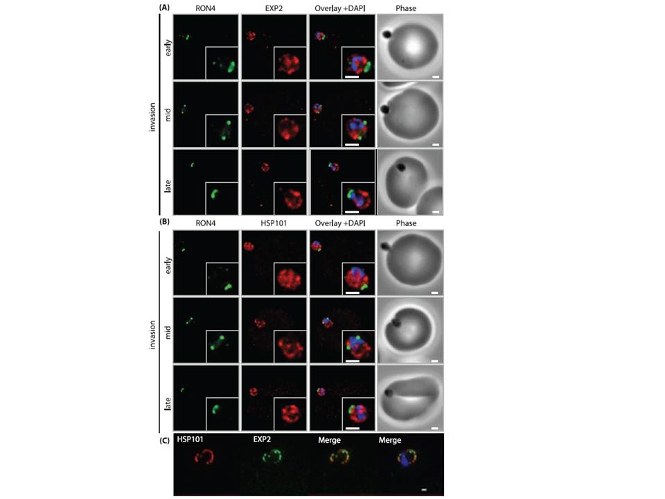
PTEX components are carried into the newly infected erythrocyte and are present at the PVM throughout intraerythrocytic development. (A and B) Time-course of merozoite invasion by widefield immunofluorescence microscopy with deconvolution (single slice shown) labelled for (A) tight junction component RON4 and PTEX component EXP2 or (B) RON4 and PTEX component HSP101. Both PTEX components are carried by the invading merozoite into the erythrocyte. Scale bar: 1μm. (C) HSP101 and EXP2 co-localise to distinct puncta at the PVM during later ring stage development. Scale bar: 1μm.
Bullen HE, Charnaud SC, Kalanon M, Riglar DT, Dekiwadia C, Kangwanrangsan N, Torii M, Tsuboi T, Baum J, Ralph SA, Cowman AF, de Koning-Ward TF, Crabb BS, Gilson PR. Biosynthesis, localisation and macromolecular arrangement of the Plasmodium falciparum translocon of exported proteins; PTEX. J Biol Chem. 2012 287(11):7871-84
Other associated proteins
| PFID | Formal Annotation |
|---|---|
| PF3D7_1116800 | heat shock protein 101 chaperone protein ClpB2 |
| PF3D7_1471100 | exported protein 2 |