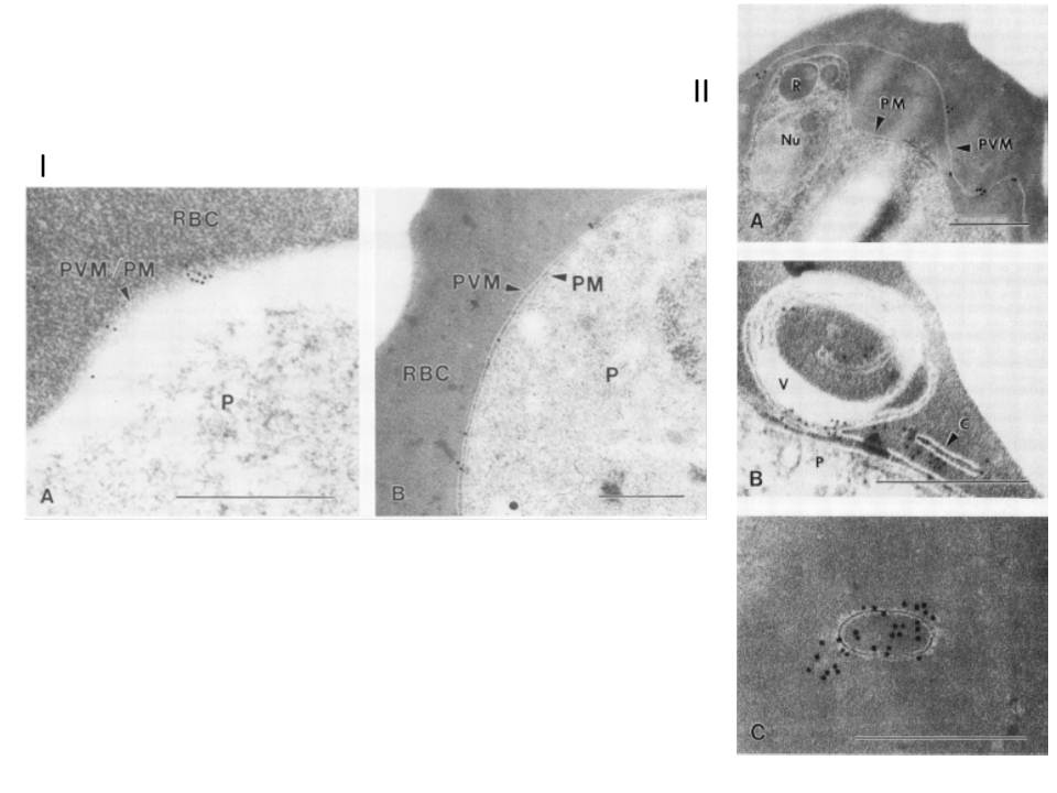
I. Immunoelectron micropgraph of P. falciparum-infected erythrocytes (FCQ-7 strain) labeled with MAb 8E7/55 and colloidal gold IgG complex. A. Schizont-infected erythrocyte showing a distinct separation of PVM from PM, with antibody labeling restricted to the vacuole membrane. Abbreviations: R, rhoptry; Nu, nucleus. (B) Membrane-bound cleft and multimembranous vesicle (V) showing antibody labeling. The parasite (P) is also shown. (C) Dense antibody labeling associated with a membrane-bound vesicle free of electron-dense material. Bars, 0.5 mm.
II. Immunoelectron micropgraph of P. falciparum-infected erythrocytes (FCQ-7 strain) labeled with MAb 8E7/55 and colloidal gold IgG complex. A. Ring-stage parasite surface labeled with gold particles (diameter, 15 nm). (B) Trophozoite-stage arasite labeled with gold particles (diameter, 10 nm). Bars, 0.5 mm. Erythrocyte (RBC) and parasite (P) are separated by both the PVM and the PM.
Kara UA, Stenzel DJ, Ingram LT, Bushell GR, Lopez JA, Kidson C. Inhibitory monoclonal antibody against a (myristylated) small-molecular-weight antigen from Plasmodium falciparum associated with the parasitophorous vacuole membrane. Infect Immun. 1988 Apr;56:903-9.