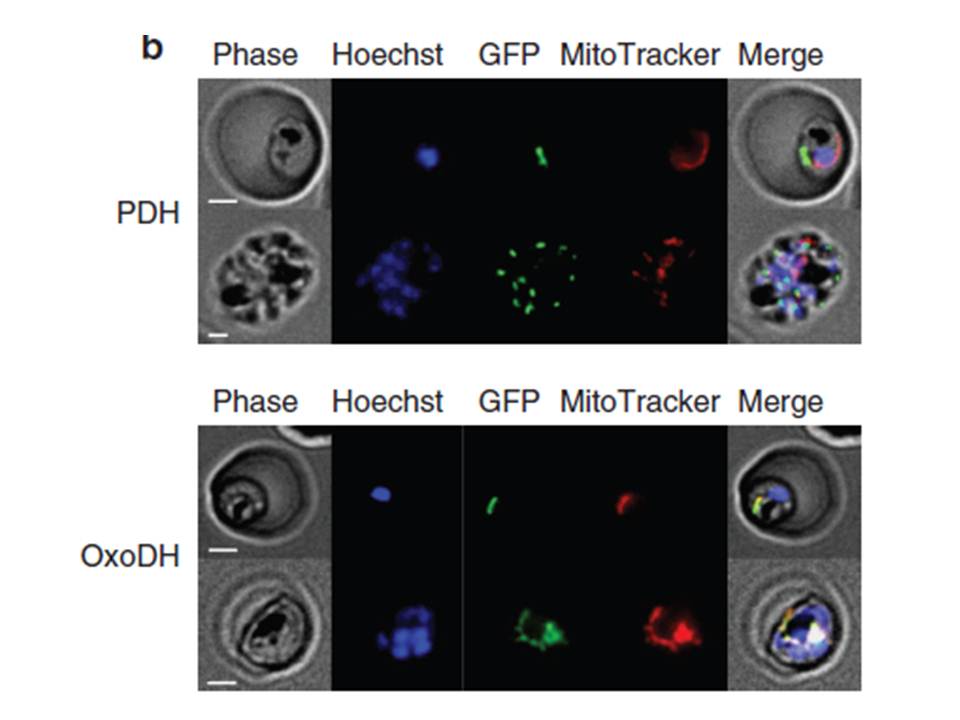
Localization of PfPDH and PfOxoDH within erythrocytes infected with 3D7 parasites. Localization of PfPDH (top panel) and PfOxoDH (bottom panel) in transgenic parasites. The left column shows phase contrast images, followed by fluorescence images of a nuclear dye (Hoechst; blue), GFP-tagged leader sequences of the target enzymes (green), a mitochondrial dye MitoTracker; red) and all four images merged together (right column). Scale bars, 2 mm. The E1-a subunit of PfPDH localizes to a discrete organelle adjacent to the mitochondrion and distinct from the nucleus, consistent with an apicoplast localization (top panel). The truncated E1 subunit of PfOxoDH, which has been predicted to have a mitochondrial localization,colocalizes with MitoTracker when fused to GFP (bottom panel), consistent with a mitochondrial localization.
Chan XW, Wrenger C, Stahl K, Bergmann B, Winterberg M, Müller IB, Saliba KJ. Chemical and genetic validation of thiamine utilization as an antimalarial drug target. Nat Commun. 2013 Jun 27;4:2060.
Other associated proteins
| PFID | Formal Annotation |
|---|---|
| PF3D7_1124500 | pyruvate dehydrogenase E1 component subunit alpha |