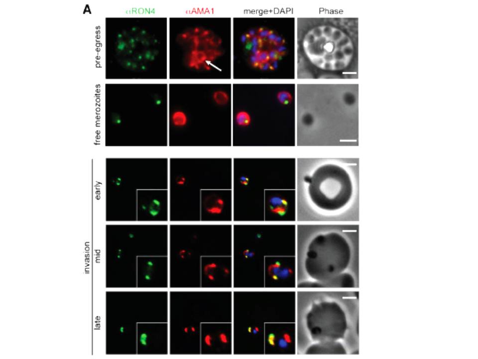
Wide field IFA time course of invasion from pre-egress through to invasion using anti-PfAMA1/PfRON4. Pre-egress and free merozoites show standard wide-field image. Invasion images show IFA with deconvolution (single slice). Scale bar = 2.0μm. In parasites fixed prior to or immediately after egress from E64-treated schizonts (ETS) a significant proportion of PfAMA1 had already been secreted onto the merozoite surface. Conventional fluorescence imaging of midinvasion merozoites revealed that PfAMA1 labeling localized directly within the plane of the ring of PfRON4 fluorescence. A proportion of PfAMA1 was also variably localized to the merozoite surface and unreleased micronemes.
Riglar DT, Richard D, Wilson DW, Boyle MJ, Dekiwadia C, Turnbull L, Angrisano F, Marapana DS, Rogers KL, Whitchurch CB, Beeson JG, Cowman AF, Ralph SA, Baum J. Super-resolution dissection of coordinated events during malaria parasite invasion of the human erythrocyte. Cell Host Microbe. 2011 9:9-20.
Other associated proteins
| PFID | Formal Annotation |
|---|---|
| PF3D7_1116000 | rhoptry neck protein 4 |