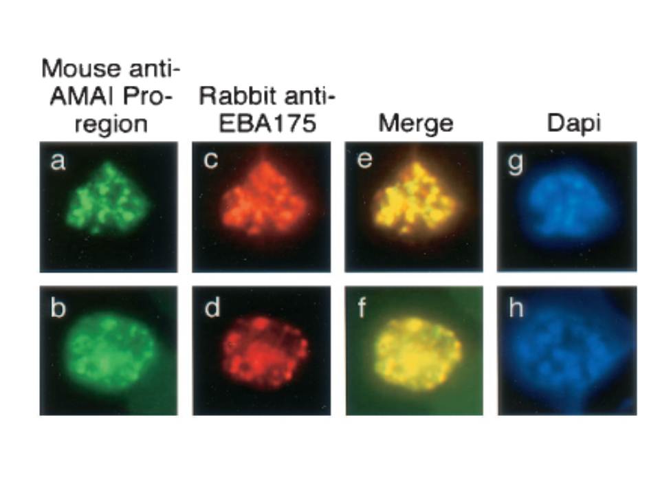
Single and dual indirect immunofluorescence images showing the same subcellular localization of AMA1 and EBA-175 in two mature schizonts of P. falciparum. (a to d) FITC-labeled MAb 5G8 against the PfAMA1 pro-region (a and b) and rhodamine-labeled anti-EBA175 (c and d). (e and f) Merged images show full-length AMA1 colocalized with EBA175. (g and h) DAPI-stained parasites included to show the stage of merozoite development. MAb 5G8, which specifically recognizes the unprocessed form of AMA1, showed a punctate pattern characteristic of the apical organelles of merozoites (a and b). Anti-EBA175 antibodies showed the same pattern (c and d), and merging of the images from the FITC and rhodamine channels suggested that these two proteins were colocalized in the same subcellular compartment, the micronemes.
Healer J, Crawford S, Ralph S, McFadden G, Cowman AF. Independent translocation of two micronemal proteins in developing Plasmodium falciparum merozoites. Infect Immun. 2002 70:5751-8.
Other associated proteins
| PFID | Formal Annotation |
|---|---|
| PF3D7_0731500 | erythrocyte binding antigen-175 |