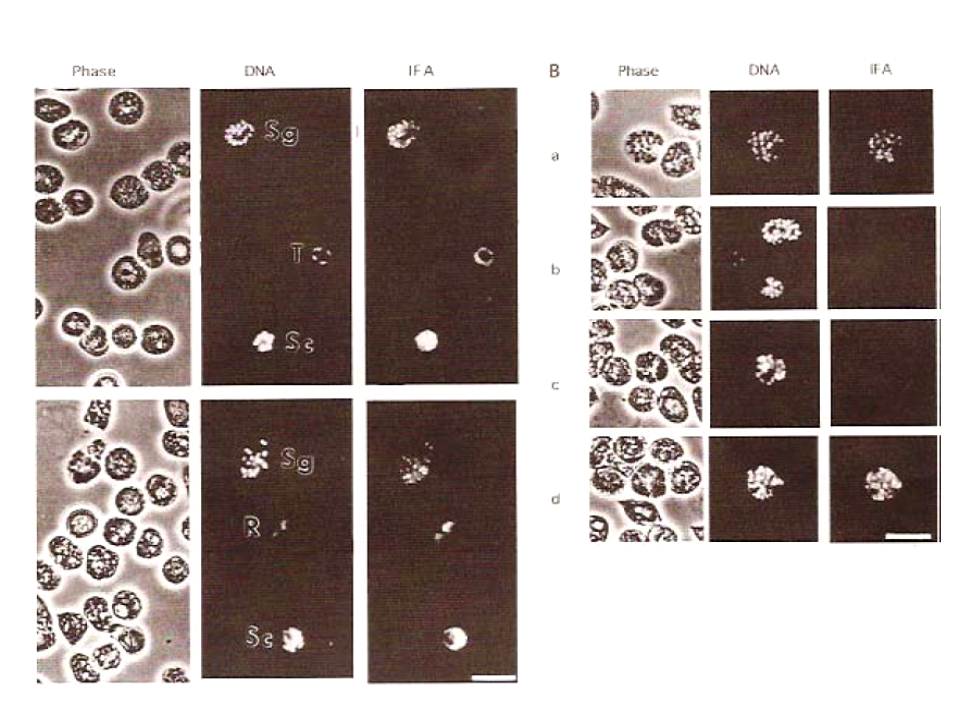
Immunofluorescence localization of Rab6 within P. falciparum-infected erythrocytes. (A) Thin films of cultured parasites were fixed in methanol and incubated with anti-Rab6i antiserum (a), or anti-Rab6i IgG fraction (b), followed by fluorescein-conjugated secondary antibody. Parasites at different stages of the intraerythrocytic cycle (R, ring; note this erythrocyte is infected with two parasites; T, trophozoite: SC, schizont; Sg, segmentor) are indicated. A phase contrast image (Phase) together with the corresponding bisbenzimide H33342 fluorescence (DNA), and immunoflourescence (IFA) images are shown for each sample. Scale bar = 10 mm. (B) lmmunofluorescence controls. Thin films of cultured parasites were fixed in methanol and incubated with anti-Rab6i antiserum (a) pre-immune serum (b) or anti-Rab6i IgG fraction (c, d), followed by fluorescein-conjugated secondary antibody. Before applying to the parasites, the anti-Rab6i IgG used in (c, d) was pre-incubated for 60 min at 23°C with either 125 ,uM peptide Rab6i (c) or 125 /tM peptide Rab6c (d). Scale bar = 5 mm.
Immunofluorescence microscopy shows that the distribution of PfRab6 changes during the final stages of parasite maturation, coalescing into multiple foci, each of which is associated with the nucleus of a forming daughter parasite.
de Castro FA, Ward GE, Jambou R, Attal G, Mayau V, Jaureguiberry G, Braun-Breton C, Chakrabarti D, Langsley G. Identification of a family of Rab G-proteins in Plasmodium falciparum and a detailed characterisation of pfrab6. Mol Biochem Parasitol. 1996 80:77-88. Copyright Elsevier