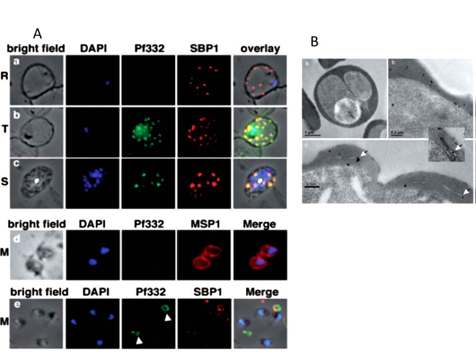
A. Localization of Pf332 in ring stage-infected erythrocytes (R) (a), trophozoite-infected erythrocytes (T) (b) and schizont-infected erythrocytes (S) (c). Each panel contains from left to right: bright field, DAPI-stained nuclei, anti-Pf322, anti-SBP1 PFE0065w and overlay of all four. Free merozoites (M) are shown in d. d is as above except that the fourth panel was labelled with anti-MSP1 PFI1475w antibodies. Panel row e contains free merozoites and material positive for both Pf332 and SBP1 suggesting Maurer’s clefts (white arrows) that were released during schizont rupture.
B. High pressure frozen and freeze substituted sections were labelled with either rabbit or mouse anti-Pf332 antibody and show the distribution of Pf332 in the red blood cell cytosol (a and b) and on Maurer’s clefts (c and inset).
Hodder AN, Maier AG, Rug M, Brown M, Hommel M, Pantic I, Puig-de-Morales-Marinkovic M, Smith B, Triglia T, Beeson J, Cowman AF. Analysis of structure and function of the giant protein Pf332 in Plasmodium falciparum. Mol Microbiol. 2009 71:48-65. Copyright John Wiley & Sons Ltd. 2010.