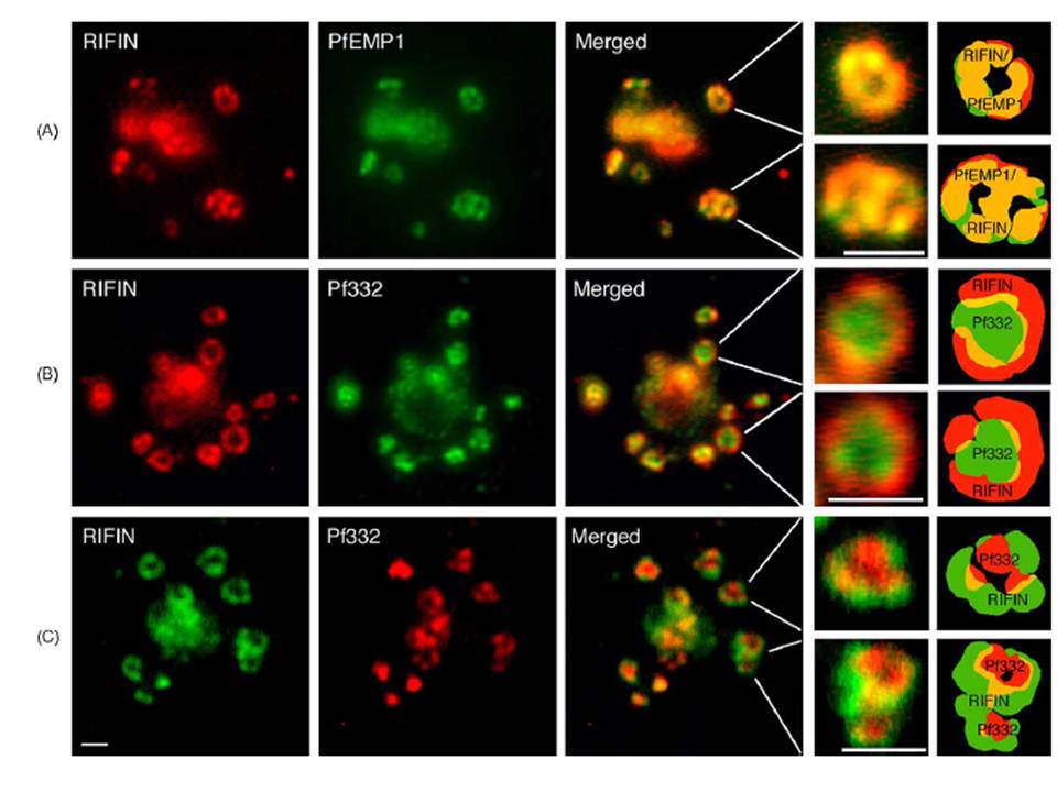
RIFIN and PfEMP1 polypeptides co-localize in the same complex in which the Maurer’s cleft antigen Pf332 is transiently co-transported. (A) Immunofluorescence staining with anti-RIFIN and anti-PfEMP1 antibodies. (B, C) Staining with anti-RIFIN and anti-Pf332 sera. Labeling of proteins was performed on air-dried monolayers of FCR3S1.2(K−) cultures at the trophozoite stage (18–24 h) as described in Section 2. Co-localization of FITC-labeled PfEMP1, Pf332 or RIFIN (green) and TRITC- or CY3-labeled RIFINS and Pf332, respectively (red), is shown in merged images (yellow). Details and illustrations show co-transport of RIFINS, PfEMP1 and Pf332 in individual large multimeric vesicles (LMV). Pf332 often occupies the center or core of the transport complex whereas RIFINS (and PfEMP1) are located in the outer rim of the assembly. Scale bar: 1 mm.
Haeggström M, Kironde F, Berzins K, Chen Q, Wahlgren M, Fernandez V. Common trafficking pathway for variant antigens destined for the surface of the Plasmodium falciparum-infected erythrocyte. Mol Biochem Parasitol. 2004 133:1-14. Copyright Elsevier 2010.
Other associated proteins
| PFID | Formal Annotation |
|---|---|
| PfEMP1 | PfEMP1 |
| RIFIN | RIFIN |