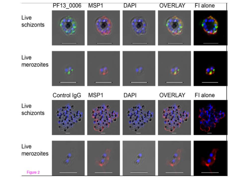
Immunofluorescence analysis of PF13_0006 expression in live asexual Plasmodium falciparum parasites. Live schizonts (first row) and merozoites (second row) of the 3D7 parasite line were analyzed by staining with anti-PF13_0006 IgG (green) and anti-MSP-1 Ab (red). As a control, live schizonts (third row) and live merozoites (fourth row) were stained with control IgG (green) and anti-MSP-1 Ab (Red). Nuclei were stained with DAPI (blue). DIC shadow-cast images with the fluorescence image superimposed in the first four coloumns and the fluorescence image (FI) alone in the last coloumn to augment the visualisation of the staining. Scale bar 5 mm. Abs stained the dividing merozoites inside the infected erythrocyte. Location of Rifin appeared to overlap with MSP-119.
Mwakalinga SB, Wang CW, Bengtsson DC, Turner L, Dinko B, Lusingu JP, Arnot DE, Sutherland CJ, Theander TG, Lavstsen T. Expression of a type B RIFIN in Plasmodium falciparum merozoites and gametes. Malar J. 2012 Dec 21;11(1):429.
Other associated proteins
| PFID | Formal Annotation |
|---|---|
| PF3D7_0930300 | merozoite surface protein 1 |